Key Takeaways
- Fluorescent labels bind specific targets, providing molecular specificity that brightfield contrast does not.
- Advantages include sensitivity, multiplexing, live-cell compatibility, and quantitative measurement.
- Photobleaching, phototoxicity, and autofluorescence are practical constraints that affect experimental design.
- System choice depends on sample type, required resolution, fluorophores, and budget.
What Is Fluorescence Microscopy?
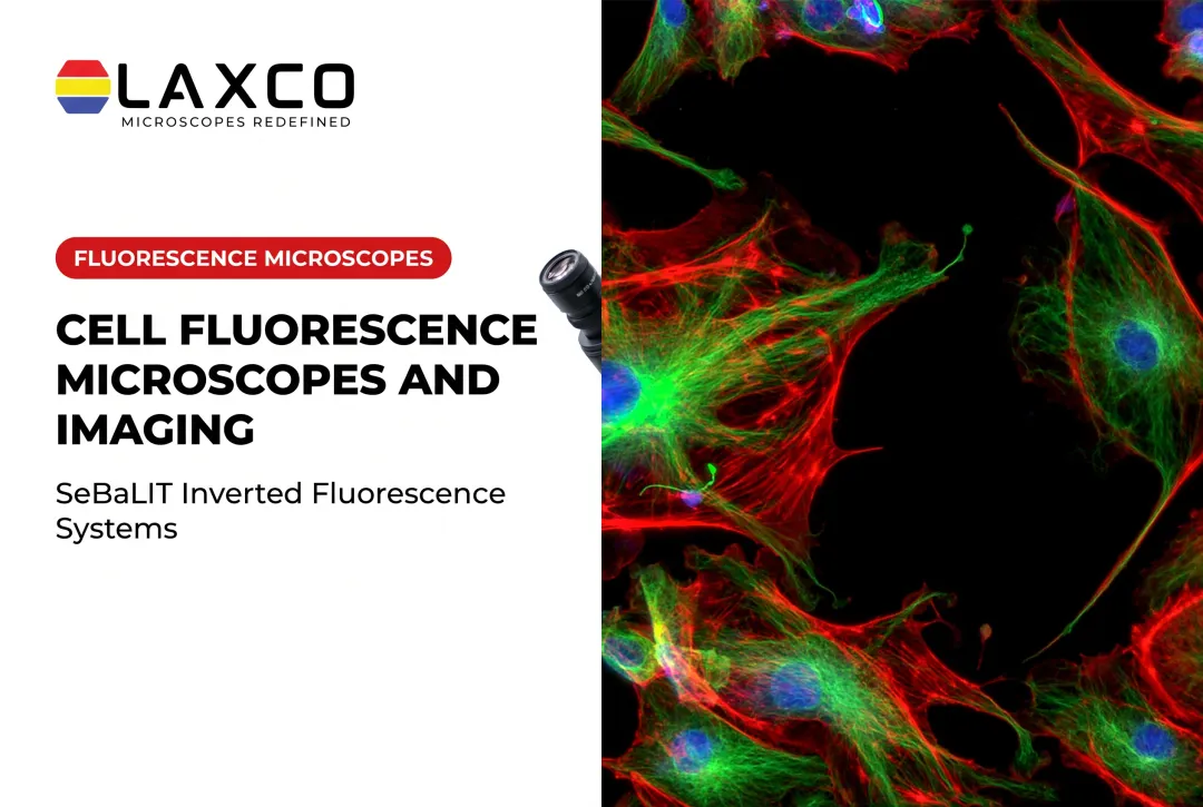
Fluorescence microscopy forms images from light emitted by fluorophores rather than from light absorbed by the sample. The microscope delivers excitation light and isolates the weaker emitted light, producing labeled structures against a dark background.
Fluorescence-based immunological labeling emerged in the early 1940s. In their original 1941 study, Coons, Creech, and Jones demonstrated that antibodies conjugated with a fluorescent compound retained their immunological specificity and could make targeted pneumococci visible under ultraviolet illumination. This work helped establish the foundation for immunofluorescence microscopy.
Fluorescence compared with brightfield:
- Brightfield contrast comes from light absorption and sample density.
- Fluorescence contrast comes from emission by labeled molecules.
- Fluorescence supports multicolor labeling of several targets in one sample.
The Principle Behind Fluorescence Microscopy
Excitation, Emission, and the Stokes Shift
A fluorophore absorbs a photon, loses some energy, and emits a lower-energy photon at a longer wavelength. The difference between the excitation and emission peaks is the Stokes shift. It ranges from a few nanometers to more than a hundred, depending on the molecule.
DAPI is a common example. It binds preferentially to AT-rich regions in the minor groove of double-stranded DNA. Its excitation maximum is around 358 nm and its emission maximum around 461 nm when bound to DNA. Its fluorescence increases substantially on binding, which produces bright nuclear staining.
The Optical Path
A fluorescence microscope separates excitation light from the weaker emitted signal:
- Light source: arc lamps, LEDs, or lasers.
- Excitation filter: selects the wavelength band that reaches the sample.
- Dichroic mirror: reflects excitation light toward the sample and transmits longer-wavelength emission.
- Emission filter: blocks residual excitation light.
- Objective and detector: collect and record the emitted signal.

In epifluorescence microscopes, the same objective illuminates the sample and collects the emitted light.
Types of Fluorescence Microscopy
| Type | Commonly used for |
|---|---|
| Widefield | Fast imaging of thin samples, with the whole field illuminated at once |
| Confocal | Optical sectioning of thicker specimens using pinhole rejection |
| Two-photon | Imaging deeper in scattering tissue |
| TIRF | Events near the coverslip surface, within about 100 nm |
| Super-resolution (STED, PALM, STORM) | Detail below the diffraction limit of roughly 0.2 µm |
Advantages of Fluorescence Microscopy
Specificity and Sensitivity
Fluorescent labels distinguish target structures from surrounding tissue. Single-molecule sensitivity is achievable under specialized conditions, but it is not the routine detection sensitivity of all fluorescence microscopes. In a 2024 Nature Nanotechnology study, Chu and colleagues demonstrated multiplexed single-molecule fluorescence detection using engineered FRET-based labels and a specialized anti-Brownian electrokinetic trapping system.
Multiplexing
Fluorophores with distinct emission spectra allow several targets to be imaged in the same sample. The number of usable channels depends on spectral overlap, filters, and detectors. Research methods using spectral unmixing and cyclic staining extend multiplexing further.
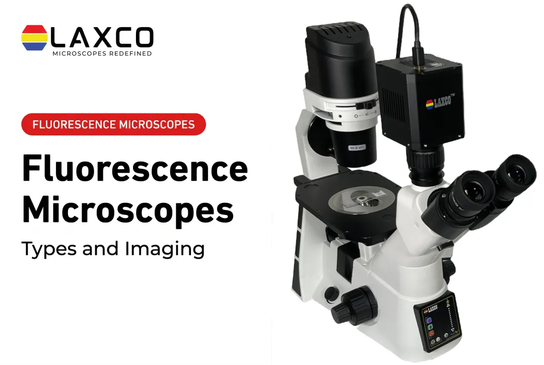
Laxco's inverted fluorescence microscopes support multichannel work:
- The SLi3Pro supports common DAPI, FITC, and TRITC fluorochromes and is field-upgradeable to fluorescence and digital imaging.
- The SLi6Pro is an inverted widefield fluorescence microscope that supports three-color multiplex imaging across approximately 390–662 nm and is compatible with more than 110 fluorochromes, including CFP, YFP, and RFP. The 110-plus figure describes dye compatibility, not simultaneous imaging of 110 markers.
Live-Cell Imaging and Quantitative Analysis
Fluorescent proteins and live-cell dyes allow dynamic processes to be observed without fixation. Examples include cell division, protein trafficking, and migration. Under controlled conditions, fluorescence intensity can be used for quantitative comparisons between samples.
Common applications include:
- Immunofluorescence for protein localization.
- Fluorescence in situ hybridization (FISH) for gene mapping.
- Drug-discovery screening.
- Biomarker studies in research samples.
Limitations to Consider
- Photobleaching: intense or prolonged illumination permanently destroys fluorophores, limiting total imaging time.
- Phototoxicity: excitation light can damage live cells or alter their normal biological behavior, even without fluorescent labels. In a 2017 Nature Methods discussion of phototoxicity, Laissue and colleagues reported changes in the proliferation and behavior of unlabeled Vero cells following blue-light exposure. The observed effects depended on the experimental illumination conditions and cell model. No single exposure dose should be treated as a universally safe or harmful threshold across all cell types and imaging systems.
- Autofluorescence: native fluorescence from molecules such as NAD(P)H and collagen adds background. Filtering and spectral unmixing reduce but may not remove it.
- Cost and training: fluorescence systems cost more and require more operator training than basic brightfield microscopes.

These limitations influence exposure settings, fluorophore selection, and sample handling.
When to Use a Fluorescence Microscope
Fluorescence microscopy is suited to questions that require identifying specific molecules:
- Protein localization by immunofluorescence.
- Gene mapping by FISH.
- Live-cell dynamics, such as division and trafficking.
- Detection of research biomarkers.
Matching the System to the Sample
Sample type, required resolution, and fluorophore panel should guide system selection. For standard three-color panels built around DAPI, FITC, and TRITC, Laxco's SLi3Pro inverted widefield fluorescence microscope supports three-color multiplex imaging with those commonly used fluorochromes.
For broader panels that extend into cyan, yellow, and far-red ranges, the SLi6Pro supports three-color multiplex fluorescence across approximately 390–662 nm and lists compatibility with more than 110 dyes. Neither model is a confocal or super-resolution system; both are inverted widefield fluorescence microscopes.
CellLive systems are designed for imaging inside CO₂ incubators. The CL11 is primarily used for brightfield confluence assessment and time-lapse monitoring. The CL22 adds phase contrast, multichannel fluorescence, Z-stack scanning, and quantitative imaging capabilities.
For separate research image-analysis requirements, Laxco's LEAP AI pairs Accuva LEAP optics with Media Cybernetics Image-Pro AI. It supports AI-assisted segmentation, measurement, batch processing, and reporting through an Image → Process → Measure → Report workflow. LEAP AI is a separate microscopy and image-analysis platform, not a built-in feature of SLiPro or CellLive instruments. Researchers considering multiple platforms should confirm workflow compatibility with Laxco.
Frequently Asked Questions
Which Laxco microscope covers DAPI, FITC, and TRITC?
The SLi3Pro supports common DAPI, FITC, and TRITC fluorochromes with SeBaLIT™ illumination. It is field-upgradeable, so fluorescence and digital imaging can be added to an existing configuration.
Which Laxco microscope suits larger fluorophore panels?
The SLi6Pro supports three-color multiplex fluorescence with an expanded approximately 390–662 nm range and is compatible with more than 110 fluorochromes, including CFP, YFP, and RFP. The compatibility figure represents the range of supported dyes, not the number of markers imaged simultaneously. Share your fluorophore list with a Laxco specialist to confirm illumination, filter, and detection compatibility for the intended experiment.
How is confocal microscopy different from widefield fluorescence?
Both use fluorescence contrast. Confocal microscopy adds pinhole rejection of out-of-focus light, enabling optical sectioning of thicker samples. Widefield microscopy is faster and simpler but does not section optically.
How can photobleaching be reduced?
Use the lowest illumination intensity and shortest exposure that give adequate signal. Sensitive cameras, photostable dyes, and antifade mounting media for fixed samples also help.