Key Takeaways
- Spatial transcriptomics keeps tissue architecture intact, so expression can be compared between tumor regions.
- Regions such as the invasive front and tertiary lymphoid structures (TLS) are studied for associations with treatment response.
- Sectioning, region selection, and imaging quality influence spatial data before sequencing begins.
- Spot-based and in-situ imaging platforms trade gene coverage against cellular resolution.
What Is Spatial Transcriptomics?
Spatial transcriptomics profiles gene expression and records the physical coordinates of that expression. The output is a map of which genes are active in which regions or cells.
The National Cancer Institute defines the tumor microenvironment as the normal cells, molecules, and blood vessels surrounding a tumor. The TME includes:
- Cancer cells, often in several molecular states within one tumor.
- Immune cells, including T cells, macrophages, and B cells.
- Cancer-associated fibroblasts (CAFs) and other stromal cells.
- Vasculature and extracellular matrix.
Limits of Bulk and Single-Cell Methods
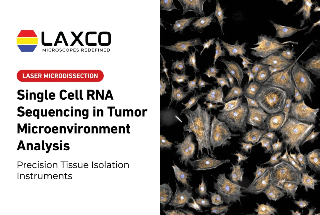
Bulk sequencing averages expression across many cells. Single-cell sequencing resolves cell states but removes cells from their tissue position. Neither method shows whether a signal originates at the tumor core or the invasive edge.
Spatial heterogeneity is the variation in cellular composition and gene expression across regions of the same tumor. Two biopsies from one tumor can differ molecularly depending on where they were taken. Spatial methods measure this variation directly.
What Spatial Transcriptomics Reveals About the TME
Spatial Organization of Cell Types
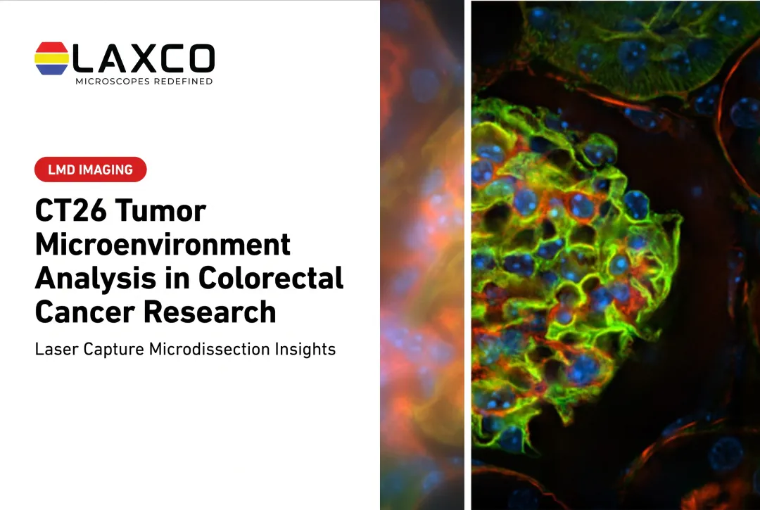
A 2025 high-resolution spatial transcriptomics study of five FFPE colorectal tumors mapped how major cell types are organized (Nature Genetics, 2025). In that study, within a 50 µm analysis window:
- CAFs clustered near tumor cells.
- Macrophages made up most of the immune compartment.
- CD4+ and CD8+ T cells were concentrated at the tumor periphery.

The invasive front and tumor core can differ in composition and expression. Tertiary lymphoid structures, which are lymph-node-like cell aggregates within tumors, have been studied as correlates of immunotherapy response.
Inferring Cell-Cell Communication
Spatial ligand-receptor analysis infers possible signaling between neighboring cells based on co-location and expression. These inferences generate hypotheses. Functional experiments are needed to confirm that a proposed interaction occurs.
Studying Treatment Response
Spatial data can be collected before and after treatment to study how tumors change. Studies in non-small cell lung cancer have examined how CAF subtypes relate to T-cell exhaustion and TLS formation in patients treated with immune checkpoint inhibitors. One multi-cohort analysis included 509 patients and related spatial and molecular features of the tumor microenvironment to immunotherapy context (PMC11866545).
In a phase 1 trial (NCT03299946) of neoadjuvant cabozantinib and nivolumab in hepatocellular carcinoma, spatial transcriptomics was used to compare responders and non-responders on a small set of quality-controlled samples (7 QC samples in the reported spatial analysis). Responder tissue showed immune-activation and B-cell signatures, whereas non-responder tissue showed metabolism and proliferation programs (Genome Medicine, 2023).
Spatial features studied as potential research biomarkers include:
- Distance to vasculature.
- Immune-cell density at tumor margins.
- CAF-tumor proximity.
Spatial Transcriptomics Platform Types
Platform choice depends on the trade-off between gene coverage and cellular resolution.
| Platform type | Resolution | Gene coverage | Commonly used for |
|---|---|---|---|
| Sequencing-based (e.g. spot-based capture) | Multi-cell spots, with higher-resolution variants available | Whole transcriptome or broad capture | Discovery studies |
| In-situ imaging | Single-cell or subcellular | Targeted gene panels | Testing defined hypotheses |

Practical considerations:
- FFPE compatibility: several current platforms support FFPE tissue, allowing archived samples to be studied when RNA quality is adequate.
- Benchmarks: sensitivity varies with tissue quality and protocol, so published comparisons may not transfer directly to other sample types.
- Specifications: vendor figures typically reflect controlled conditions.
Whole-transcriptome methods suit exploratory questions. Targeted panels suit validation of defined hypotheses, such as TLS composition or CAF states. Discovery and targeted designs are complementary rather than exclusive: many studies begin with broad spatial profiling and follow with targeted panels or orthogonal assays on the same or matched samples.
Sample Preparation and Region Isolation
Poor sectioning, damaged morphology, or imprecise region selection add noise before sequencing starts.
Isolating Defined Regions
Laser capture microdissection (LCM) isolates selected regions from tissue sections for downstream molecular analysis. Examples include an invasive front, a TLS, or a tumor-stroma boundary. LCM complements spatial transcriptomics when a region identified on a spatial map needs targeted follow-up. LCM provides region-based collection of material; it does not itself generate a continuous tissue-wide transcriptomic map.
Laxco's Accuva Cellect Laser Capture Microdissection System provides:
- An infrared (IR) laser designed for film capture of single cells or small numbers of cells.
- An ultraviolet (UV) laser designed for cutting dense structures, such as bone or plant tissue.
- A five-step software workflow with visual quality control and documentation.
- Documented support for FFPE and fresh-frozen tissue, H&E-stained and LCM-optimized stains, immunofluorescence, unstained samples, and cell culture.
RNA purification, reverse transcription, library preparation, and sequencing are separate downstream laboratory procedures, not functions of the instrument. Captured material can proceed to those steps using methods validated for the collection device, extraction chemistry, and assay.
Image-Based Region Selection
Manual identification of regions of interest can vary between users, especially in complex tumor tissue. Laxco's LEAP AI is a separate imaging and analysis platform that combines Accuva LEAP optics with Media Cybernetics Image-Pro AI. It is not built into Accuva Cellect and does not produce spatial transcriptomics data. Its capabilities include:
- Pre-trained deep-learning models for AI-assisted segmentation.
- Batch processing with no-code protocols.
- Image stitching, alignment, and enhancement.
- Measurement and reporting of segmented regions.
Segmentation should be validated for each stain and tissue type. It supports consistent region selection but does not replace validated spatial-sequencing chemistry, does not automatically transfer selections to Accuva Cellect, and does not control the Cellect laser.
Challenges and Future Directions
- Cost and throughput: these vary by platform, panel breadth, and tissue area.
- Computation: normalization, cell segmentation, and noise reduction require dedicated analysis pipelines.
- Depth: some platforms detect fewer transcripts per cell, so studies often pair spatial data with bulk RNA-seq or proteomics.
Large collaborative programs, such as the NCI Human Tumor Atlas Network, are building multi-assay atlases of tumors from precancerous lesions to advanced disease.
Frequently Asked Questions
How is spatial transcriptomics different from single-cell RNA sequencing?
Single-cell sequencing resolves individual cell states but loses tissue location during dissociation. Spatial transcriptomics preserves location, sometimes with lower per-cell resolution. Many studies integrate the two data types computationally.
Can archived FFPE samples be used for spatial transcriptomics?
Yes, on FFPE-compatible platforms, provided RNA quality is adequate. Fixation, storage time, and section handling affect results, so quality checks are advisable before committing samples.
When is laser capture microdissection useful alongside spatial methods?
LCM is useful when a region identified on a spatial map or stained section needs targeted molecular follow-up. Isolated material can be analyzed by qPCR or low-input RNA sequencing as separate downstream steps. Accuva Cellect performs the microdissection and collection; it does not run the molecular assays itself.
Which sample types does Accuva Cellect support?
Laxco lists FFPE and fresh-frozen tissue, H&E-stained and LCM-optimized stained sections, immunofluorescence, unstained samples, and cell culture. Confirm compatibility with your downstream assay during planning.