Key Takeaways
- Organoids can be generated from pluripotent stem cells, adult tissue-derived cells, or patient-derived tumor material, producing 3D structures that reproduce selected features of the source tissue.
- They are used in cancer research, drug screening, disease modeling, and developmental biology.
- Patient-derived tumor organoids have been compared with clinical treatment responses in published studies.
- Organoids typically lack vasculature and immune components and often reflect fetal or early developmental stages.
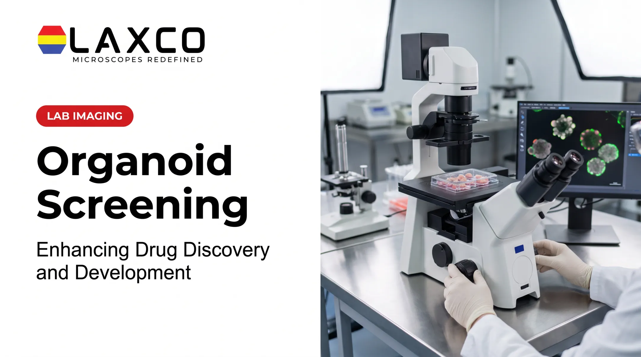
- Microscopy, image analysis, and laser capture microdissection (LCM) support the measurement and sampling of organoid cultures.
What Are Organoids?
An organoid is a 3D cellular model that self-organizes into structures reproducing selected architectural, cellular, and functional characteristics of a tissue or organ.
Organoids can be established from several cellular sources:
- Pluripotent stem cells: embryonic stem cells or induced pluripotent stem cells (iPSCs) can be directed toward particular tissue lineages and used to study organ development and differentiation.
- Adult tissue-derived stem and progenitor cells: cells obtained from primary tissue can form organoids that retain selected characteristics of their tissue of origin.
- Patient-derived tumor cells: suitable tumor specimens can be used to establish patient-derived tumor organoids that preserve some of the genetic, histological, and molecular features of the source tumor.
Organoid culture conditions vary by source and model. Tissue-specific growth factors, signaling molecules, and extracellular matrices help support survival, expansion, or differentiation.
Animal-derived basement membrane extracts remain common in organoid culture, while defined and synthetic matrix systems are being developed to improve reproducibility and reduce batch-to-batch variation.
The National Cancer Institute's definition of organoids includes structures grown from tumor cells or stem cells.
Organoids and Spheroids
A spheroid is a 3D aggregate of cells that may not develop organ-specific structure. An organoid self-organizes into tissue-like architecture with distinct cell types. Both are 3D models, but they answer different experimental questions.
Organoid Maturity
Organoids have been generated for intestine, liver, kidney, lung, brain, and other tissues. Maturity varies by tissue and protocol. Brain organoids, for example, reproduce features of developing brain tissue but do not form a functional adult brain.
Biomedical Applications of Organoids
Cancer Research and Drug Response
Patient-derived tumor organoids (PDOs) can retain genetic and histological features of the original tumor. A 2018 study in Science established PDOs from patients with metastatic colorectal and gastroesophageal cancer enrolled in clinical trials (Vlachogiannis et al., 2018). The authors compared organoid drug responses with patients' clinical responses. In that cohort, the reported sensitivity was 100% and specificity was 93% for predicting response.
These results apply to the drugs, cancer types, and patients studied. Predictive performance has varied in other studies and treatment settings.
Drug Discovery and Toxicity Studies
Organoids can provide human-derived evidence about biological responses and potential toxicity during preclinical drug development. Their relevance depends on the organoid model, compound, measured endpoint, and validation of the experimental method.
The FDA Modernization Act 2.0, enacted through the Food and Drug Omnibus Reform Act of 2022, expanded the statutory recognition of nonanimal approaches as possible sources of nonclinical evidence for drug development. This created greater flexibility to use suitable cell-based assays and other scientifically supported methods alongside or, where appropriate, instead of certain animal studies.
On September 21, 2026, the U.S. Food and Drug Administration announced a regulatory update concerning alternatives to animal testing. The update clarifies the role of scientifically valid nonanimal methods, including human-cell-based systems, organ-on-chip platforms, and computational approaches, while retaining the need for evidence appropriate to the product's safety evaluation.
These developments do not mean that animal testing is universally unnecessary or that organoid data automatically satisfy regulatory requirements. The acceptability of a nonanimal method depends on the intended use, scientific validity, and applicable regulatory expectations.
Organoid models should therefore be presented as an expanding component of biomedical research and drug-development testing, not as universally validated replacements for all in vivo studies.
Disease Modeling and Developmental Biology
Organoids allow researchers to study human developmental processes that are difficult to observe in vivo. For example, studies published in 2016, including Garcez et al. in Science, reported that Zika virus infection reduced the growth of human brain organoids. This work helped link infection of neural progenitor cells to developmental brain defects.
Personalized Medicine Research
Patient-derived organoids are being studied as functional tests of individual drug response. Cystic fibrosis research has used patient-derived organoid swelling assays to assess CFTR modulator response. These remain research applications unless validated for clinical use.
Imaging and Isolation Methods for Organoid Studies
Organoid studies generate morphological, molecular, and functional data. Several methods support these measurements.
Fluorescence Microscopy
Fluorescence microscopy visualizes labeled cell types, proteins, and structures within organoids. Multichannel imaging can show several markers in the same sample. Many organoids are optically thick or scattering, which can limit imaging depth with widefield methods. Sections, clearing, or optical sectioning methods may be needed for deeper structures.
Image Analysis
High-throughput organoid experiments can produce large numbers of images. Automated segmentation and measurement can reduce manual workload and some sources of analyst-to-analyst variability, depending on the model and validation dataset.
Laser Capture Microdissection
Organoids can contain several cell types and differentiation states. LCM isolates selected cells or regions from organoid sections for downstream DNA, RNA, or protein analysis. This keeps the molecular sample linked to the morphology that guided its selection.
Laxco Systems for Organoid Research
Laxco instruments can support several steps of an organoid workflow:
- SLi6Pro inverted fluorescence microscope: patented SeBaLIT™ illumination covering 390–662 nm and compatibility with more than 110 fluorochromes. It supports multichannel fluorescence of cultured samples.
- CellLive: live-cell imaging systems that operate inside CO₂ incubators, with scheduled imaging, motorized autofocus, and remote monitoring. Laxco lists stem cell and organoid research among their applications.
- LEAP AI: combines Accuva LEAP optics with Media Cybernetics Image-Pro AI for pre-trained deep-learning segmentation, measurement, batch processing, and reporting.
- Accuva Cellect: a laser capture microdissection system with an infrared laser for capture of single cells or small numbers of cells and a UV laser for cutting dense structures. Laxco lists FFPE and fresh-frozen tissue, several staining types, and cell culture as supported samples.
Laxco supplies these instruments with application training and technical support. It does not perform organoid culture or analysis as a laboratory service.

Recent Advances in Organoid Research
Organoid research is moving beyond relatively simple epithelial or stem-cell-derived structures toward models that incorporate additional cell types, improved tissue organization, and more detailed molecular characterization.
Three developments are particularly relevant to current biomedical research.
1. Vascularized Organoid Models
A major limitation of many conventional organoids is the absence of mature vascular networks. Without appropriate transport structures, diffusion becomes increasingly important as organoid size and complexity increase.
Researchers are developing models that incorporate endothelial cells and tissue-specific vascular structures. In 2025, investigators reported lung and intestinal organoid models in which coordinated development of different cellular lineages supported the formation of specialized vascular components.
The National Institutes of Health highlighted this vascularized-organoid research in August 2025.
These developments may improve the study of tissue formation and cell interactions, but the presence of vascular structures does not automatically mean an organoid has the fully functional circulation of an intact organ.
2. Organoid-Immune Cell Co-Cultures
Many conventional organoid cultures lack the immune components necessary to reproduce inflammatory signaling and tissue-immune interactions.
Co-culture approaches introduce selected immune-cell populations into organoid experiments. These systems can help researchers investigate immune responses, inflammatory disease mechanisms, and interactions between tumor cells and immune populations.
A 2025 Cell Reports review of organoid models of immune-organ interactions discusses applications in mucosal immunity, inflammation, and oncology.
Such models remain experimentally simplified and require validation of immune-cell composition, function, and persistence.
3. More Detailed Spatial and Molecular Characterization
Improved imaging, sequencing, and computational approaches are making it possible to examine not only which cell types exist in an organoid but also how their molecular states relate to internal tissue organization.
In 2026, Nucera and colleagues published a spatial transcriptomics benchmarking study across eight stem-cell-derived organoid models. The researchers examined regional molecular profiling, multi-organoid sample placement, and limitations affecting RNA capture efficiency.
This research illustrates why organoid characterization increasingly depends on reproducible sample preparation, spatially informed measurements, and suitable quality-control methods.
For laboratories, the practical implication is that no single imaging or molecular method fully characterizes an organoid. Microscopy, quantitative image analysis, single-cell methods, spatial profiling, and targeted tissue isolation can provide complementary information when chosen according to the research question.
Current Limitations
Organoids are useful models, but they differ from intact organs in several ways:
- Limited mature vasculature. Many conventional organoid cultures lack a mature, perfused vascular network, so oxygen and nutrient transport largely depends on diffusion. This can limit growth and contribute to central stress or necrosis in larger organoids. Some engineered organoid models incorporate endothelial cells or vascular structures, although these do not automatically reproduce the fully functional circulation of an intact organ.
- Developmental immaturity. Many organoids resemble fetal or early developmental tissue, which can limit modeling of adult-onset disease.
- Incomplete cellular composition. Many conventional organoid protocols lack some of the immune, vascular, stromal, or other supporting cell populations found in intact tissues. Co-culture and engineered systems can introduce selected additional cell types, but the resulting models still may not reproduce the full cellular complexity of an organ.
- Variability. Organoids can vary in size, shape, and composition between batches and wells, which affects reproducibility.
Current work aims to address these gaps through co-culture with immune or vascular cells, organ-on-chip systems, and more defined matrices.
Frequently Asked Questions
Which Laxco system suits long-term organoid imaging?
CellLive systems are designed for continuous imaging inside CO₂ incubators, with scheduled time-lapse imaging and remote monitoring. The CL22 adds phase contrast, multichannel fluorescence, and Z-stack scanning for more detailed observation.
How can I image several markers in the same organoid?
Multichannel fluorescence on an inverted microscope suits stained organoids and cultures. The SLi6Pro uses SeBaLIT illumination across 390–662 nm with more than 110 compatible fluorochromes. Thick organoids may need clearing or sectioning for internal detail.
Can I isolate specific cells from organoid sections?
Yes. Accuva Cellect isolates selected cells or regions from sections, with an infrared laser for single cells or small groups and a UV laser for dense structures. This links molecular data to organoid structure.
Can automated analysis replace manual organoid measurements?
Automated segmentation can measure size, number, and shape consistently across many wells. LEAP AI uses pre-trained deep-learning segmentation, but results should be validated against manual measurements for each organoid type.
What should I compare before requesting a quote?
Define your organoid types, imaging frequency, required contrast methods, and whether you need regional isolation. A Laxco specialist can then recommend CellLive, SLi6Pro, LEAP AI, Accuva Cellect, or a combined workflow.