Key Takeaways
- Light and electron microscopy use different imaging mechanisms and have different resolution and sample-preparation requirements; digital image capture and analysis can be incorporated into either approach.
- Fluorescence and confocal methods reveal labeled structures that brightfield imaging cannot distinguish.
- Numerical aperture (NA) and wavelength determine the resolution of a light microscope.
- Image analysis software converts images into quantitative measurements.
- System choice depends on sample type, resolution needs, budget, and analysis workflow.
What Is Microscopy and Imaging?
Microscopes can be used for direct observation through eyepieces, digital image acquisition, or both. Digital imaging adds cameras or other detectors and software that capture, store, measure, analyze, and share images.
Digital microscopy is not a separate optical category equivalent to light or electron microscopy. Instead, it describes how microscope images are acquired, displayed, and processed. Digital acquisition can be incorporated into multiple microscopy technologies.
| Aspect | Direct eyepiece observation | Digital image acquisition |
|---|---|---|
| Output | Real-time optical view through the eyepieces | Captured images or video displayed and stored electronically |
| Analysis | Visual interpretation and manual measurements where supported | Software-assisted visualization, segmentation, measurement, and reporting |
| Sharing | Direct viewing or use of separately attached recording equipment | Digital transfer, remote review, and electronic archiving |
| Measurement | Calibrated eyepiece reticles or other manual measurement methods | Calibrated image-based measurements using suitable software |

Microscopy is used in life sciences research, clinical laboratories, manufacturing quality control, education, and materials science.
Types of Microscopy
Light Microscopy
Light microscopy uses visible light and glass lenses. Common contrast methods include:
- Brightfield: transmitted light for stained or naturally colored specimens.
- Phase contrast: converts differences in optical path into contrast, which suits transparent live cells.
- Darkfield: shows scattered light against a dark background.
- Polarized light: increases contrast in birefringent materials, such as crystals, minerals, and fibers.
Fluorescence and Confocal Microscopy
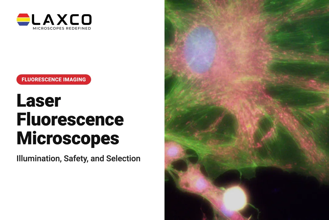
- Fluorescence: labeled probes absorb light at one wavelength and emit at a longer wavelength. Targets appear bright against a dark background.
- Confocal: a pinhole rejects out-of-focus light, producing optical sections that can be combined into 3D reconstructions.

Electron Microscopy
Electron microscopes use electron beams instead of light, which allows much finer resolution.
- Scanning electron microscopy (SEM) scans a beam across a surface to show topography and composition.
- Transmission electron microscopy (TEM) passes electrons through thin sections to show internal structure.
Electron microscopy generally requires specialized sample preparation and does not image living cells.
Digital and AI-Assisted Microscopy
Digital microscopes integrate a camera, computer, and display. Image analysis software can apply deep-learning models to segment cells or features, measure them, and generate reports. Automated analysis can reduce differences between operators, provided it is validated on representative images.
How Imaging Works
In a typical digital light-microscopy workflow, the sample is illuminated, objectives form a magnified optical image, and a camera captures the image for display, storage, or software analysis.
Electron microscopy uses a different imaging process involving an electron beam and appropriate detectors. The acquisition and signal-detection methods therefore depend on the microscopy technology being used.
Resolution depends on wavelength and numerical aperture. NA is defined as n × sin(α), where n is the refractive index of the medium and α is half the objective's angular aperture. Higher NA gives finer resolution.
Conventional light microscopy is limited by diffraction to roughly 200 nm laterally. Electron microscopy reaches far finer resolution, although biological sample preparation often limits the detail that can be achieved in practice.
Image analysis turns pixels into data through segmentation, measurement, and reporting.
Laser Capture Microdissection
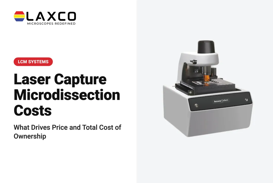
Some systems combine imaging with sample isolation. Laser capture microdissection (LCM) uses a laser to isolate selected cells or tissue regions from a slide for genetic or molecular analysis. The researcher identifies the target under the microscope before capture.
Applications
Life Sciences and Clinical Laboratories
Microscopy supports research on cell structure, protein localization, and tissue morphology. In clinical laboratories, stained specimens remain central to several diagnostic workflows:
- Malaria: blood film microscopy is the standard method for confirming malaria (CDC).
- Tuberculosis: sputum smear microscopy for acid-fast bacilli is one of the core diagnostic methods (CDC).
- Hematology: peripheral blood smears show abnormal cell counts and shapes.
- Pathology: many cancer diagnoses rely on examining cell and tissue appearance.

Cancer Research
Tumor samples contain mixed cell types. LCM allows researchers to isolate tumor, precancerous, and normal cells separately for gene expression or mutation analysis.
Industrial Quality Control
Microscopes are used to inspect semiconductor wafers, flat panel displays, and fiber optic connectors. Fiber connector end faces are inspected for contamination and defects before connection.
Education
Digital microscopes allow students to share views, capture images, and prepare lab reports together.
Choosing a Microscopy System
- Sample type: live cells, fixed tissue, industrial materials, and fiber connectors require different optics.
- Resolution: light microscopy suits most cellular work, while electron microscopy is needed for nanoscale structure.
- Contrast methods: confirm support for brightfield, phase contrast, darkfield, polarization, or fluorescence as needed.
- Analysis: decide whether automated segmentation and batch processing are needed.
- Support: consider whether optics, cameras, and software are supported by one supplier.
Laxco Microscopy Systems
- Upright microscopes: the LMC-4000 Series supports brightfield, darkfield, phase contrast, fluorescence, and metallurgical methods.
- SLi3Pro and SLi6Pro: inverted fluorescence microscopes with SeBaLIT™ illumination. The SLi6Pro covers 390–662 nm and more than 110 fluorochromes.
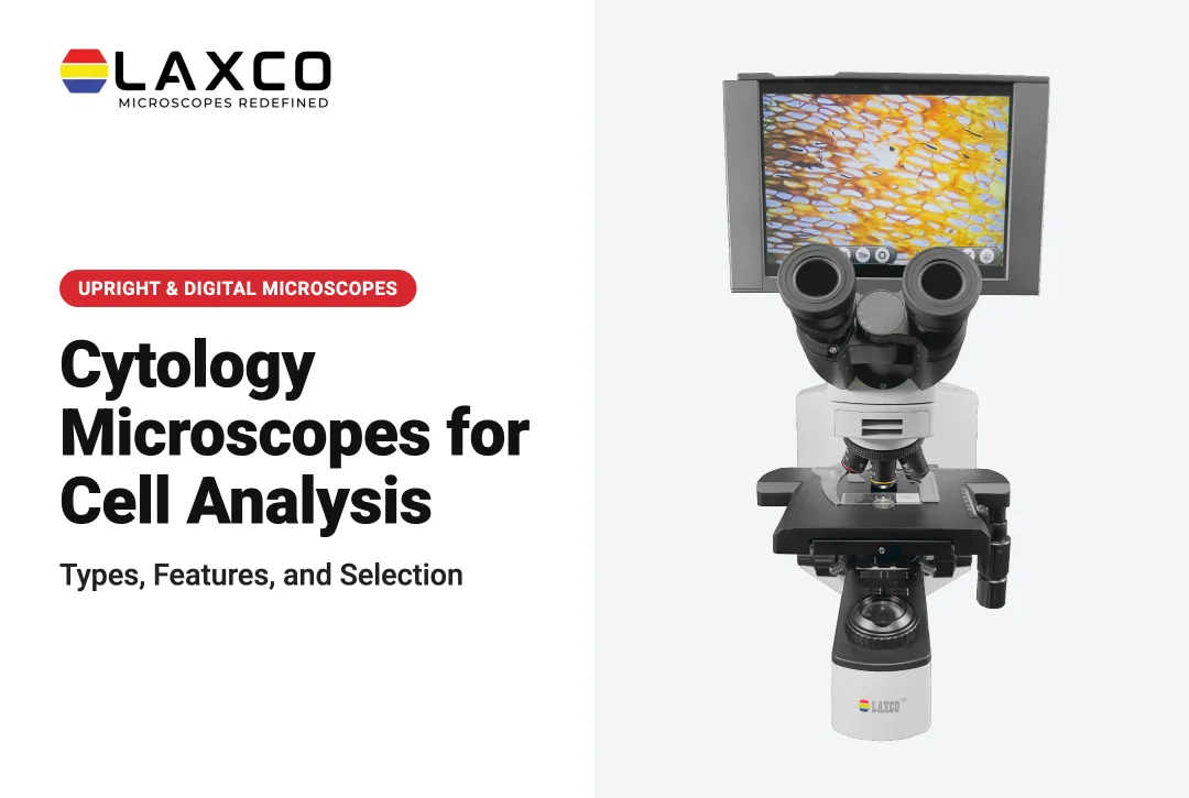
- SeBa digital microscopes: integrate camera, computer, and monitor for teaching, collaboration, and documentation.
- LEAP AI: pairs Accuva LEAP optics with Media Cybernetics Image-Pro AI in an Image, Process, Measure, Report workflow.
- Accuva Cellect: a laser capture microdissection system with IR and UV lasers.
- LWI Series and FML Series: inspection microscopes for wafers and flat panel displays, and field microscopes for fiber connectors in 100X, 160X, and 200X versions.
Frequently Asked Questions
Which microscopy type should I choose for my samples?
Choose by sample and question. Light microscopy suits most cell and tissue work, fluorescence adds labeled targets, confocal adds optical sectioning, and electron microscopy reaches nanoscale structure. Also consider live versus fixed samples and analysis needs.
What limits the resolution of a light microscope?
Diffraction limits conventional light microscopy to roughly 200 nm laterally. Achieved resolution depends on wavelength, objective numerical aperture, sample preparation, and focus. Higher magnification without higher numerical aperture does not reveal more detail.
Which clinical tests still rely on microscopy?
Microscopy remains important in clinical laboratory workflows, including malaria blood-film examination, acid-fast bacilli (AFB) smear examination during tuberculosis evaluation, peripheral blood-smear review, and histopathology.
AFB smear microscopy can help identify potentially infectious specimens but does not independently confirm tuberculosis, because acid-fast organisms may include mycobacteria other than Mycobacterium tuberculosis. The CDC's clinical and laboratory guidance explains the role and limitations of smear microscopy.
Appropriately configured Laxco clinical microscopes may support relevant specimen observation workflows. Laboratories must confirm instrument configuration, intended use, and validation requirements for the specific clinical application. Diagnosis and result interpretation remain the responsibility of qualified laboratory professionals.
How does AI-assisted analysis change microscopy workflows?
Deep-learning tools automate segmentation, measurement, and reporting across many images, which reduces manual outlining. LEAP AI pairs Accuva LEAP optics with Image-Pro AI for this. Results should be validated against manually annotated images.
Which Laxco microscopes support fluorescence imaging?
The SLi3Pro and SLi6Pro are inverted fluorescence microscopes with SeBaLIT illumination, and the LMC-4000 Series upright microscopes support fluorescence among other methods. The SLi6Pro covers 390–662 nm with more than 110 compatible fluorochromes.