Key Takeaways
- LCM isolates selected cells from tissue sections under microscopic guidance, reducing contributions from neighboring cell types.
- Direct-lysis workflows can reduce RNA losses by avoiding a separate purification step, but the collection method, lysis chemistry, and reverse-transcription reaction must be compatible and validated for very small inputs.
- Low copy numbers increase technical noise, so controls and replicate design are essential.
- Minimizing time between capture and lysis helps protect RNA integrity.
- Oncology, developmental biology, and neuroscience studies use this workflow to profile defined cell populations.
What Is Laser Capture Microdissection?
LCM isolates specific cells or regions from a tissue section while the operator views it under a microscope. Instead of inferring which cells contributed to a bulk signal, the researcher selects the cells directly.
Common laser microdissection approaches include:
- IR thermoplastic-film capture: an infrared laser activates a film-coated capture surface that adheres to selected cells or tissue regions. The material is recovered with the capture surface. This is a specific film-based collection method rather than a universal mechanism for every laser microdissection instrument.
- UV laser cutting: a focused ultraviolet laser cuts around selected cells or tissue regions. The separated material is recovered using the collection mechanism supported by the instrument.
- Laser-assisted transfer or catapulting: some UV-based microdissection systems use an additional laser pulse to propel cut material into a collection device. This is a collection mechanism used in particular systems, not a third universal laser architecture alongside IR and UV.
Tissue Preparation
- Fresh-frozen tissue generally offers better RNA preservation, while formalin-fixed paraffin-embedded (FFPE) tissue often offers well-preserved morphology and access to archived samples. Both can support suitable molecular assays, but direct RT-PCR without RNA purification must be validated for the preservation method. A protocol developed for rapidly stained frozen tissue should not automatically be applied to FFPE sections.
- Thin sections, commonly in the 5–10 µm range, are used for single-cell capture.
- Stains should allow cell identification while preserving RNA. Options include H&E, immunofluorescence, and LCM-optimized stains.
How Single-Cell RT-PCR Works
Reverse-transcription PCR (RT-PCR) converts RNA into complementary DNA (cDNA) and amplifies selected sequences using PCR. Conventional RT-PCR can be used to detect or characterize expressed transcripts, including through nested PCR, but it does not automatically provide reliable quantitative expression measurements. Quantitative comparisons require an appropriately validated RT-qPCR or other quantitative assay, suitable normalization, and controls. With single-cell input, RNA loss, incomplete reverse transcription, and low transcript copy number can substantially affect detection.
Why low input changes the approach:
- RNA content varies between cell types and between individual cells.
- Expression of a single gene can vary widely from cell to cell.
- At low copy numbers, random sampling increases variability and can cause dropout.
Direct lysis without purification:
- Confirm collection compatibility. Determine whether the LCM platform supports a validated on-cap lysis and reverse-transcription method or requires transfer of captured material into a separate reaction vessel.
- Choose compatible chemistry. Use RNase-controlled consumables and a lysis formulation validated for the reverse-transcription enzyme. Detergents, fixation-related compounds, and other carryover substances may inhibit downstream reactions.
- Minimize transfers where validated. Carry out reverse transcription directly in the collection vessel or on the capture surface only when the workflow and consumables support that procedure.
- Control contamination. Include no-template, no-reverse-transcriptase, and appropriate positive controls. Assess whether the workflow also requires genomic-DNA removal or transcript-specific primer design.
- Validate amplification. Use nested PCR or preamplification only when appropriate for the target and assay, and assess detection sensitivity, dropout, and amplification bias.
Published Example: Single-Cell RT-PCR Without RNA Purification
Keays et al. (2005) studied individual CD38-positive plasma cells isolated from rapidly immunostained frozen human brain sections. The researchers compared two approaches for reverse transcription of unpurified cell lysates. Their more successful method performed reverse transcription directly on detergent-solubilized material on the capture-cap surface, followed by nested PCR.
Under the study's conditions, immunoglobulin heavy- and light-chain transcripts were detected in approximately two-thirds of individually isolated plasma cells. This demonstrates the feasibility of purification-free, on-cap single-cell RT-PCR in a validated experimental workflow. The detection rate, reagent chemistry, and collection method should not be treated as universal specifications or as a validated protocol for every current LCM instrument.
Integrated Workflow
- Section and stain the tissue.
- Identify the target cells or region.
- Capture the target and initiate the validated lysis or stabilization workflow promptly, either on the capture surface or after transfer to a compatible reaction vessel.
- Reverse-transcribe the lysate.
- Amplify with appropriate controls.
Time and RNA Stability
RNA in tissue sections can degrade during microdissection as sections rehydrate and endogenous RNases become active. A 2008 study reported two methods that stabilized RNA during microdissection for up to 90 minutes: working under an argon atmosphere and using ethanol-based staining solutions (Clément-Ziza et al., 2008). Validate the acceptable time window under your own laboratory conditions.
Contamination Control
- Clean work surfaces and use RNase-controlled consumables.
- Run no-reverse-transcriptase controls to detect genomic DNA.
- Design primers that span exon junctions where possible.
- Document selected cells with images for later review.
Research Applications
Oncology: isolating tumor cells from adjacent stroma reveals expression differences that bulk samples can obscure, including genes related to drug metabolism and response.
Developmental biology: tracking gene expression in specific epithelial or mesenchymal populations during tissue formation requires precise, time-point-specific capture.
Neuroscience: pooling small numbers of neurons or glial cells of one type allows expression comparisons between neighboring cell populations.
Limitations and Troubleshooting
Limitations:
- Detection: transcripts not converted during reverse transcription are not detected.
- Amplification bias: low starting copy numbers increase variability.
- Limited multiplexing: without preamplification, only a few targets can be measured per cell.
- Fixation trade-offs: frozen tissue protects RNA but offers less morphological detail than FFPE.
| Problem | Likely cause | Possible solution |
|---|---|---|
| Degraded RNA | Delay between capture and lysis | Shorten the interval, or use stabilizing conditions |
| Low cDNA yield | Inefficient reverse transcription | Reduce reaction volume and use fresh RNase inhibitor |
| Inconsistent amplification | Low copy number | Add validated preamplification or pool cells |
| Signal in no-RT control | Genomic DNA contamination | Use exon-spanning primers or DNase treatment |
Choosing an LCM System for Single-Cell Work
- Capture method: suitable for single cells and small groups.
- Sample compatibility: fresh-frozen, FFPE, and the stains you plan to use.
- Collection: caps or tubes compatible with direct lysis.
- Software: documentation of selected cells and quality control.
Laxco Accuva Cellect for Single-Cell RT-PCR Workflows
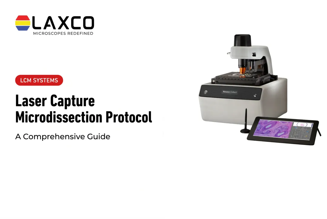
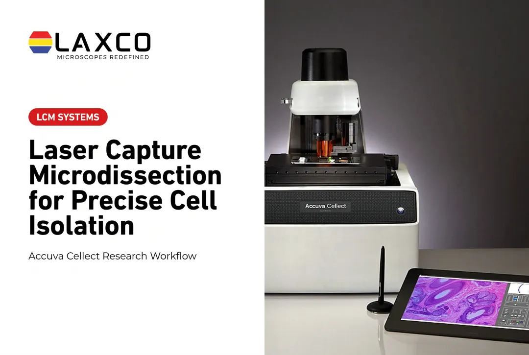
Laxco's Accuva Cellect Laser Capture Microdissection System supports microgenomics workflows that include RT-PCR:
- Infrared (IR) laser: designed for capture of single cells or small numbers of cells.
- UV laser: designed for cutting dense structures, such as bone or plant tissue.
- Sample preparation: Laxco lists FFPE and fresh-frozen tissue, H&E-stained and LCM-optimized stains, immunofluorescence, unstained samples, and cell culture.
- Collection: compatible with CapSure LCM caps.
- Software workflow: a five-step workflow with visual quality control.
Laxco also offers LEAP AI, a separate microscopy and image-analysis platform combining Accuva LEAP optics with Media Cybernetics Image-Pro AI. Its segmentation capabilities support microscopy image analysis, but Laxco does not document LEAP AI as an integrated target-selection or laser-capture feature of Accuva Cellect. Any use of separately generated image annotations to guide microdissection would require a compatible, validated workflow.

Accuva Cellect is a research instrument. Laxco's Cellect Grant Assistance Program provides Grant Language Kits for NIH, NSF, HHMI, and DOD proposals.
Frequently Asked Questions
Why skip RNA purification for single-cell RT-PCR?
Separate RNA purification can cause substantial sample loss when working with the very small amount of RNA available from individual cells. Validated direct-lysis methods can reduce handling and recovery losses, allowing reverse transcription from unpurified lysate. However, this approach requires compatible lysis chemistry and collection consumables, and inhibitors or genomic DNA may affect amplification. Keays et al. (2005) demonstrated one method involving reverse transcription on the capture-cap surface.
How many cells should I pool for low-abundance transcripts?
The appropriate input depends on RNA abundance, cell type, and assay sensitivity. Single-cell RT-PCR can detect transcripts from individual cells, but low-copy targets may be missed because of sampling and reverse-transcription variability. Pooling matched cells can improve detection by increasing input, but it sacrifices true single-cell resolution and can mask cell-to-cell differences. Use pilot experiments to choose between individual-cell and pooled-cell analysis based on the research question.
Which controls should every single-cell RT-PCR run include?
Include no-template controls, no-reverse-transcriptase controls to detect genomic DNA, and positive controls with known expression. Exon-spanning primers further reduce the risk of amplifying genomic DNA from lysed cells.
Can Accuva Cellect capture individual cells for RT-PCR?
Laxco describes the infrared laser in Accuva Cellect as designed to capture single cells or small cell populations. Captured material may be used in downstream RT-PCR workflows. However, compatibility with direct lysis, on-cap reverse transcription, or purification-free RT-PCR must be confirmed for the selected collection cap, reagents, and protocol. The instrument's single-cell capture capability does not, by itself, establish that every direct RT-PCR chemistry is supported.
Can image analysis help identify target cells before capture?
Yes. Validated image-analysis and segmentation methods can help researchers identify candidate cells or tissue regions. Results should be reviewed against tissue morphology and the requirements of the LCM workflow. Laxco's LEAP AI combines Accuva LEAP optics with Media Cybernetics Image-Pro AI, but it is a separate microscopy and image-analysis platform, not a documented integrated Accuva Cellect target-selection or capture function.