Key Takeaways
- Single-cell isolation separates individual cells for molecular or functional analysis.
- Single-cell isolation approaches include FACS, single-cell microfluidic capture, laser capture microdissection (LCM), micromanipulation, and limiting dilution. Conventional MACS primarily enriches cell populations before further isolation or analysis.
- Suspension-based methods require dissociation, while LCM works directly on tissue sections.
- Method choice depends on sample type, throughput, viability needs, and downstream assay.
- Isolation quality strongly affects scRNA-seq data quality.
Why Single-Cell Isolation Matters
Bulk analysis averages signals across many cells. Rare cells, such as a tumor cell with a resistance mutation or an uncommon stem cell, may not be detectable in that average.
Single-cell analysis can reveal:
- Cell-to-cell variation in gene expression
- Rare mutations in specific cells
- Functional differences between cells that look similar
Single-cell isolation is used in cancer research, developmental biology, immunology, and drug discovery. Contamination from neighboring cells, mechanical stress, and cell damage can bias results, so the isolation step needs careful control.
Single-Cell Isolation Techniques
Fluorescence-Activated Cell Sorting (FACS)
FACS labels cells with fluorescent antibodies or dyes, measures them as they pass one by one through lasers, and deflects selected cells into collection tubes or plates.
- Best for: dissociated suspensions requiring multi-parameter, high-speed sorting
- Strengths: high throughput, multiple markers, deposition of single cells into plates
- Limitations: requires suspensions and sufficient starting cells, can stress fragile cells, needs trained operators
Magnetic-Activated Cell Sorting (MACS)
MACS uses antibody-conjugated magnetic beads to retain labeled cells in a magnetic field while unlabeled cells pass through. Conventional MACS is primarily a population-enrichment method: it increases the proportion of target cells in a mixed sample rather than depositing one verified single cell per well. Laboratories often use MACS upstream of single-cell isolation or analysis steps (for example FACS, microfluidic capture, or plate-based workflows) when starting material is abundant and a marker-defined subpopulation must be concentrated first.
- Best for: rapid enrichment before further processing
- Strengths: simple, gentle, and cost-effective
- Limitations: separates populations rather than isolating individual cells, and depends on antibody specificity
Microfluidics
Microfluidic devices separate or trap cells in microscale channels, either by physical properties such as size and deformability or by antibody capture. Droplet-based microfluidics also encapsulates single cells for barcoding in scRNA-seq.
- Best for: rare cells in small sample volumes, such as circulating tumor cells, and high-throughput single-cell capture
- Strengths: low reagent volumes and scalable throughput
- Limitations: requires dissociated cells, devices can clog, and setup needs expertise
Laser Capture Microdissection (LCM)
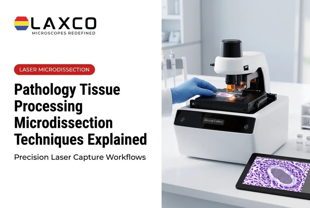
LCM uses a microscope-guided laser to isolate selected cells or regions directly from tissue sections.
- How it differs: works on intact sections, so each cell's location and surrounding morphology are documented.
- Best for: tumor microenvironment studies, spatial biology, and heterogeneous tissue.
- Strengths: selection by morphology or staining, single-cell precision, no dissociation required.
- Limitations: lower throughput than high-speed sorting, operator training required, and sample-preparation compatibility constraints. Fixed or frozen sections are generally unsuitable for culture; specialized live-cell LCM workflows can recover viable cells for some applications.

Micromanipulation and Limiting Dilution
- Micromanipulation: a micropipette, operated manually or robotically under a microscope, picks individual live cells.
- Limiting dilution: cells are diluted across wells according to a probability distribution in which some wells receive no cells, some receive one cell, and some receive more than one.
Both approaches are used for clonal cell line development and hybridoma work. Micromanipulation is labor-intensive when many clones are required. Limiting dilution alone does not guarantee monoclonality: because some wells can still receive more than one cell, laboratories typically repeat dilution rounds and apply additional confirmation steps (imaging, growth kinetics, or molecular assays) before treating a line as monoclonal. Guidance on confirming monoclonality is summarized in the literature (for example PMC12417753).
Single-Cell Isolation for scRNA-seq
Damaged cells release ambient RNA, which adds background to gene expression data. scRNA-seq isolation pathways differ by starting material:
Suspension pathway (dissociated cells):
- Dissociate tissue into a single-cell suspension.
- Isolate or enrich target cells (FACS, microfluidic capture, or related methods).
- Capture and barcode cells using droplet or well-based platforms.
- Sequence and analyze the data.
Tissue-section pathway (histologically defined cells): LCM selects cells or regions from intact sections while retaining spatial context. Low-input library methods can profile RNA from LCM material; for example, Foley et al. (2019) combined LCM with Smart-3SEQ on FFPE tissue (PMC6836736). Droplet-based scRNA-seq kits are built for intact single-cell suspensions and may not accept extracted LCM RNA as input - confirm kit requirements before planning the assay.

Careful isolation supports gene expression profiling, mutation detection, lineage tracing, and combined protein-RNA analysis.
How to Choose a Method
Consider:
- Sample source: dissociated suspension or intact tissue section
- Throughput: a few rare cells or thousands of cells
- Spatial context: whether tissue location must be recorded
- Budget and equipment: available instruments, consumables, and expertise
- Downstream application: sequencing, PCR, proteomics, or culture
- Cell fragility: whether cells tolerate sorting or dissociation
Start by distinguishing the primary goal: viable cells for culture, molecular profiling from suspensions, or histologically defined cells from intact tissue. Throughput, purity, viability, and downstream compatibility then trade off against each other; identify the requirement that cannot be compromised and choose accordingly.
Checks Before Finalizing
- Spatial context: FACS and MACS require dissociation, which removes tissue architecture.
- Contamination: control neighboring cells and ambient RNA.
- Full workflow cost: include extraction, amplification, and library preparation.
- Input requirements: confirm that the method meets your assay's input needs.
Laxco Accuva Cellect
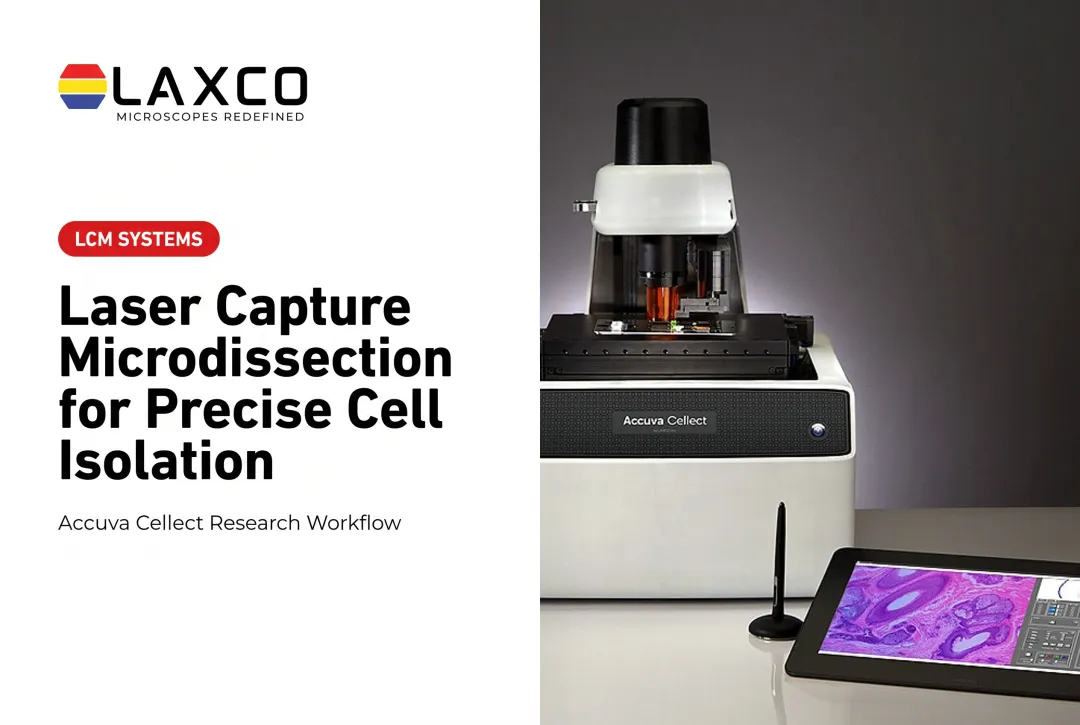
Laxco's Accuva Cellect Laser Capture Microdissection System isolates cells directly from tissue sections:
- Infrared (IR) laser: designed for capture of single cells or small numbers of cells.
- UV laser: designed for cutting dense structures, such as bone or plant tissue.
- Sample preparation: Laxco lists FFPE and fresh-frozen tissue, H&E-stained and LCM-optimized stains, immunofluorescence, unstained samples, and cell culture.
- Software workflow: a five-step workflow with visual quality control.
Laxco lists training and 48-hour response times for Accuva Cellect. Its Cellect Grant Assistance Program offers Grant Language Kits for NIH, NSF, HHMI, and DOD proposals.
Frequently Asked Questions
How do I choose a single-cell isolation method?
Match the method to sample form and goal. Use FACS when dissociated suspensions need multi-parameter sorting and plate deposition. Use single-cell microfluidic capture for high-throughput barcoding from suspensions or rare cells in small volumes. Use conventional MACS when the priority is gentle population enrichment before a true single-cell step. Use LCM when cells must be selected from intact tissue by morphology or location. Then weigh throughput, viability, spatial context, and the input requirements of the downstream assay.
When should I use LCM instead of FACS?
Use LCM when cells must be selected from intact tissue by location or morphology. Use FACS when working with dissociated suspensions and selecting by markers at high throughput. LCM also records where each cell came from.
Can LCM-isolated cells be cultured afterwards?
Most LCM workflows use fixed or frozen sections and collect cells for molecular analysis rather than culture. Specialized live-cell LCM approaches can recover viable cells in some settings (PubMed 23698319). When post-isolation culture and high viability are primary requirements, FACS deposition and micromanipulation are generally more suitable.
Which sample types does Accuva Cellect support?
Laxco lists FFPE and fresh-frozen tissue, H&E-stained and LCM-optimized stained sections, immunofluorescence, unstained samples, and cell culture. The infrared laser is designed for single cells or small numbers of cells.
Does Laxco help with funding for LCM equipment?
Yes. The Cellect Grant Assistance Program provides Grant Language Kits with technical specifications and a 15-minute Grant Fit consultation for NIH, NSF, HHMI, and DOD proposals that include Accuva Cellect.