Key Takeaways
- CT26 is a syngeneic mouse model used to study colorectal tumor-immune interactions in immunocompetent hosts.
- The TME includes immune infiltrates, stromal cells, vasculature, and signaling molecules.
- Region-specific analysis can reveal spatial differences that bulk measurements average together.
- Prespecified region-of-interest criteria and contamination checks improve comparability across samples.
- CT26 findings require careful translation, because mouse and human tumor immune composition differ.
What Is CT26 Tumor Microenvironment Analysis?
CT26 is a murine colon carcinoma cell line originally established from a chemically induced tumor in BALB/c mice following exposure to N-nitroso-N-methylurethane. CT26.WT is a clone derived from this parental line and is available as ATCC CRL-2638. When implanted into appropriately matched, immunocompetent BALB/c mice, CT26 cells form a syngeneic tumor model that allows researchers to investigate interactions between malignant cells and a functioning host immune system.
CT26 is a transplantable experimental tumor model rather than a direct recreation of human colorectal cancer. Its microenvironment can vary with implantation site, cell preparation, and host conditions. For example, subcutaneous CT26 tumors do not reproduce the anatomical environment of tumors growing within the colon.
The TME includes malignant tumor cells and non-malignant components around them. TME analysis in this model examines those non-malignant components:
- Immune infiltrates: T cells, macrophages, dendritic cells, and NK cells.
- Stromal cells: fibroblasts and cancer-associated fibroblasts (CAFs).
- Vascular structures: endothelial cells and tumor-associated vessels.
- Extracellular matrix and signaling molecules: cytokines, chemokines, and growth factors.

Common analytical methods include:
- Flow cytometry: quantifies immune-cell populations in dissociated tumors.
- Immunohistochemistry and immunofluorescence: visualize protein expression and cell location in tissue sections.
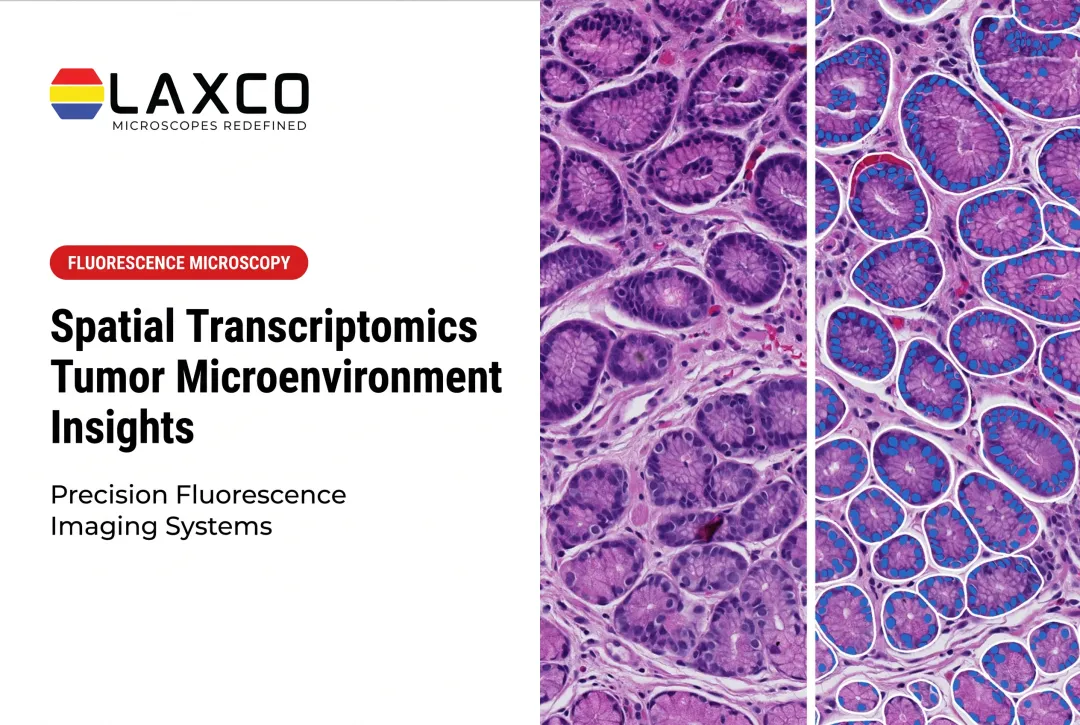
- Spatial transcriptomics: maps gene expression within tissue sections.
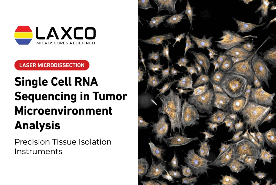
- Laser capture microdissection (LCM) with expression profiling: isolates defined regions for molecular analysis.
These methods answer different questions, and many studies combine more than one.
Why CT26 TME Analysis Matters in Colorectal Cancer Research
TME analysis in CT26 models helps researchers investigate how immunotherapies and experimental treatments interact with the tumor immune microenvironment. In a 2021 study, Sato and colleagues compared CT26 with Colon 26, another BALB/c-derived murine colon carcinoma model. Under the study's experimental conditions, CT26 tumors showed greater infiltration by several immune populations, including CD8+ T cells, dendritic cells, and natural killer cells. The researchers also reported differences in PD-1-positive CD8+ T-cell populations and found that CT26 tumors were more sensitive to anti-PD-1 treatment than Colon 26 tumors.
In this comparison, CT26 displayed a relatively immune-inflamed, or "hot," tumor microenvironment. The researchers investigated differences in Wnt-pathway signaling as a possible contributor to the contrasting immune phenotypes, but this proposed mechanism should not be treated as universally established. Immune infiltration and therapeutic response may vary with implantation site, experimental conditions, and tumor preparation. The study therefore does not establish that every CT26 tumor will display the same immune profile or respond similarly to anti-PD-1 therapy.
These model systems are used to:
- Study immune-evasion mechanisms, including regulatory T-cell-mediated suppression.
- Investigate markers associated with immunotherapy response in the model.
- Test checkpoint inhibitors and combination regimens preclinically.
- Compare immune composition between the tumor core, invasive margin, and stroma.
CT26 does not reproduce human colorectal cancer immune composition exactly. Differences in myeloid and lymphoid populations between mouse models and human tumors mean findings should be translated cautiously.
A Stepwise CT26 TME Analysis Workflow
Step 1: Define the research objective. Decide whether the study addresses immune infiltration, stromal remodeling, angiogenesis, or drug response. This choice determines the analytical methods and sampling plan.
Step 2: Harvest and prepare tumors. Collect tumors at a predefined size or time point. Consistent timing and fixation or freezing conditions improve reproducibility across cohorts.
Step 3: Identify and isolate regions of interest. Stained sections and images are used to distinguish the tumor core, invasive margin, and stromal zones. LCM can then isolate these regions before molecular analysis.
Step 4: Apply molecular or imaging analysis. Isolated regions can be analyzed by qPCR or RNA-seq, while intact sections can be analyzed by multiplex immunofluorescence. Flow cytometry requires dissociated tissue and does not retain location.
Step 5: Interpret cellular and spatial patterns. Pooled qPCR or RNA-seq from isolated regions provides region-level profiles, not a continuous spatial map. Imaging of intact sections is a separate analysis. Expression and cell-type distributions can still be related back to tumor architecture, and immune-excluded regions, stromal barriers, and vascular patterns can be compared between conditions.
Step 6: Validate findings. Confirm results across biological replicates and with orthogonal methods before drawing conclusions about treatment effects.
Common Pitfalls in CT26 TME Analysis
- Cross-contamination between compartments. Manual dissection between tumor and stroma can include unintended cells, which affects molecular readouts.
- Inconsistent region-of-interest selection. Without prespecified criteria, samples from different animals may not be comparable.
- Insufficient imaging resolution. Low resolution can miss immune-cell clustering patterns relevant to the research question.
Record these parameters before starting:
- Model identity and passage history.
- Tumor size, harvest day, and implantation site.
- Animal strain, sex, age, and housing conditions.
- Treatment schedule and controls.
- Sampling plan across animals and tumors.
- Section thickness and staining method.
- Region-of-interest rules and number of regions per tumor.
- Quality metrics for RNA or protein recovery.
- Orthogonal validation methods.
- Dissection order and contamination checks.
Laxco Tools for Region-Specific CT26 Analysis
Researchers who need molecular data from defined tumor regions may consider LCM. Laxco's Accuva Cellect Laser Capture Microdissection System provides microscopy-guided target selection:
- An infrared (IR) laser designed for film capture of single cells or small numbers of cells, such as sparse immune infiltrates.
- An ultraviolet (UV) laser designed for cutting dense structures, such as bone or plant tissue.
- A five-step software workflow with visual quality control and documentation.
- Documented support for FFPE and fresh-frozen sections and several staining approaches.
For microscopy image analysis of tumor sections, Laxco's LEAP AI combines Accuva LEAP optics with Media Cybernetics Image-Pro AI. It supports AI-assisted segmentation, measurement, and batch processing through configurable workflows, including no-code options. Segmentation results should be validated for the tissue type, staining method, and cell populations being examined.
LEAP AI is a separate imaging and image-analysis platform, not a built-in Accuva Cellect feature or a documented automated controller for its laser-capture workflow. It should not be described as automatically transferring segmented cells or regions to Cellect for capture. Researchers planning to use both platforms must confirm image registration, sample handling, and cross-platform workflow compatibility.
Laxco does not have published CT26-specific validation data comparing tumor core, margin, and stroma. Researchers should establish region-specific performance in pilot experiments. The Cellect Grant Assistance Program provides technical documentation for grant applications that include LCM.
Frequently Asked Questions
Why is CT26 used for colorectal cancer TME studies?
CT26 is syngeneic to immunocompetent BALB/c mice, so tumor-immune interactions can be studied in an intact immune system. Xenograft models in immunodeficient mice do not allow this. As a mouse model, CT26 still carries species and implantation-site caveats when findings are translated to human colorectal cancer.
Can LCM be combined with RNA-seq for CT26 studies?
Yes. LCM-isolated regions can be analyzed by RNA-seq or qPCR using low-input methods. These measurements are pooled profiles from the captured material, not single-cell profiles or a continuous spatial map. Pilot experiments help establish how much tissue area each region requires.
Which sample types does Accuva Cellect support?
Laxco lists FFPE and fresh-frozen tissue, H&E-stained and LCM-optimized stained sections, immunofluorescence, unstained samples, and cell culture. Confirm compatibility with your downstream assay during planning.
How can variability between CT26 sections be reduced?
Standardize tumor harvest, fixation, and sectioning. Prespecify region-of-interest criteria before dissection, and use consistent, validated image-analysis settings across samples.