Key Takeaways
- Spatial transcriptomics maps RNA expression, often across thousands of genes.
- Spatial proteomics maps proteins, receptor localization, and some post-translational modifications.
- RNA abundance does not always predict protein abundance or activity in the same region.
- Both modalities support discovery and validation. Spatial transcriptomics reveals gene-expression programs, while spatial proteomics provides protein-level measurements, including localization and selected modifications when supported by the assay.
- Integrated studies have measured both modalities on the same tissue section.
Comparison at a Glance
| Factor | Spatial transcriptomics | Spatial proteomics |
|---|---|---|
| Molecular target | RNA transcripts | Proteins and their localization; selected modifications with suitable assays |
| Typical coverage | Targeted gene panels to whole-transcriptome measurements, depending on platform | Defined protein panels in antibody-based imaging; broader protein identification through suitable LC-MS/MS workflows |
| Resolution | Multi-cell regions to subcellular localization, depending on method | Cellular or subcellular imaging, or region-associated measurements after tissue isolation |
| Sample types | Fresh-frozen and FFPE, where supported by the assay | Fresh-frozen and FFPE, depending on staining, protein recovery, and analytical requirements |
| Common use | Mapping gene-expression programs, cellular states, and spatial organization | Mapping protein abundance and localization, examining selected signaling markers, or identifying proteins from defined tissue regions |

What Is Spatial Transcriptomics?
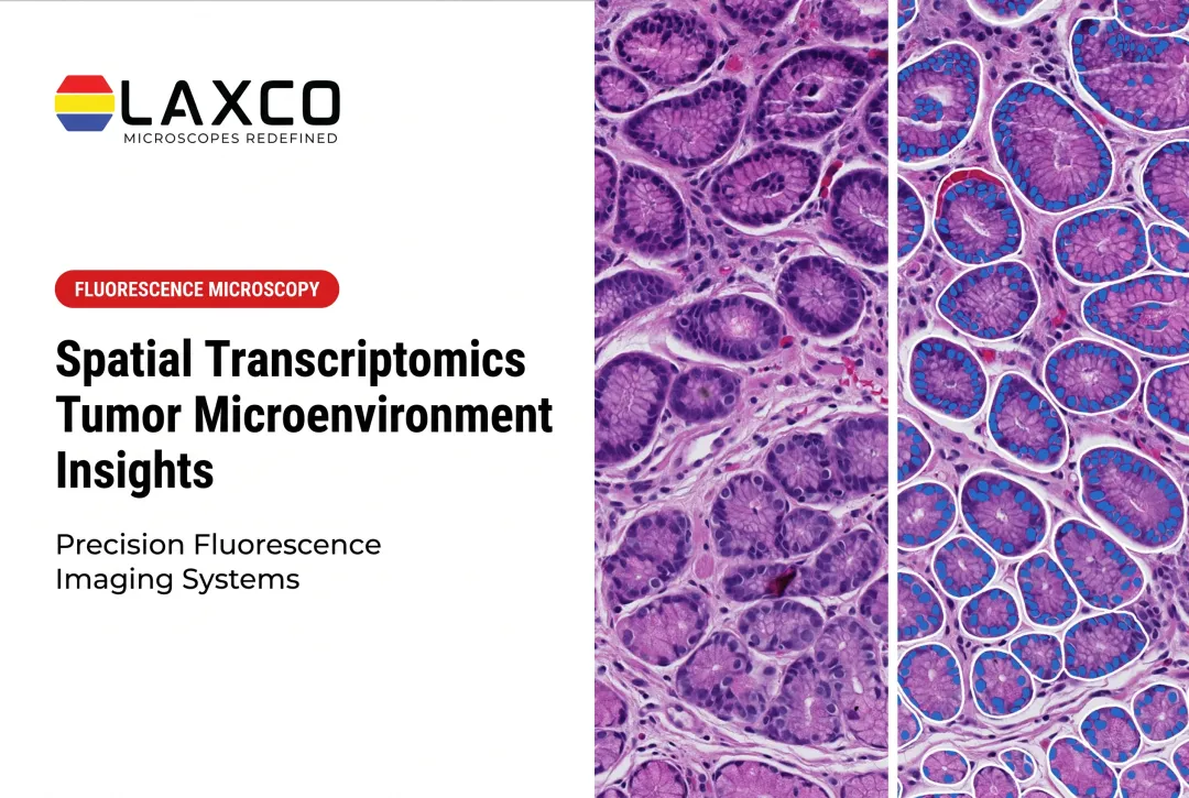
Spatial transcriptomics measures gene expression in intact tissue sections and assigns each measurement a position. This allows researchers to ask which genes are expressed in a specific region, and next to which cell types.
Methods fall into two main groups:
- Sequencing-based methods capture transcripts on barcoded surfaces and map reads back to tissue positions. Resolution ranges from multi-cell spots to near single-cell features.
- Imaging-based methods detect transcripts in situ through repeated hybridization or in situ sequencing. They usually measure a targeted gene panel at subcellular resolution.
Common applications include mapping the tumor microenvironment, tracing expression gradients in developing tissue, and distinguishing cell types across brain regions.
What Is Spatial Proteomics?
Spatial proteomics measures proteins while retaining tissue context. Protein measurements can show receptor localization, protein presence, and, with suitable reagents, post-translational modifications that RNA data cannot reveal.
Main method categories include:
- Antibody-based imaging: cyclic immunofluorescence, imaging mass cytometry, and multiplexed ion beam imaging.
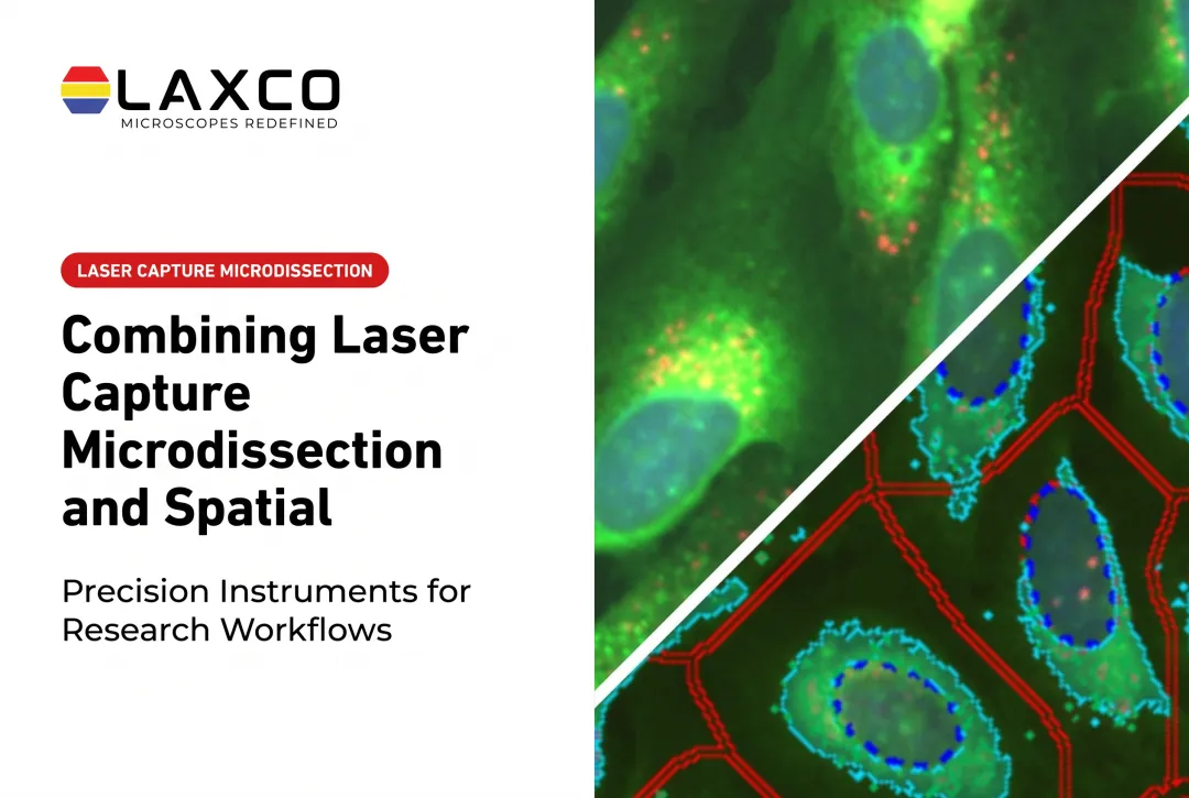
- Mass spectrometry of selected regions: cells or regions are isolated from tissue, often by laser capture microdissection (LCM), and analyzed by mass spectrometry.
Nature Methods named spatial proteomics its Method of the Year for 2024 (Nature Methods, 2024).
Antibody-based spatial proteomics uses labeled antibodies to detect selected proteins while preserving their location within tissue architecture. Different methods achieve multiplexing in different ways: cyclic immunofluorescence uses successive imaging rounds, while imaging mass cytometry and multiplexed ion beam imaging distinguish metal-labeled antibody signals using mass-based detection.
The number of measurable markers, spatial resolution, tissue compatibility, and analytical requirements depend on the specific method and platform. These imaging approaches differ from laser capture microdissection followed by LC-MS/MS, which isolates selected cells or tissue regions for separate protein analysis rather than generating a continuous spatial protein map.
Choosing Between the Two
Five factors usually guide the decision:
- Research question: whether the study needs RNA-expression patterns, protein abundance or localization, selected protein modifications, or broader molecular discovery.
- Resolution: region-level, cell-level, or subcellular.
- Sample type: fresh-frozen or FFPE.
- Budget and access: instrument availability and per-sample cost.
- Multiplexing: thousands of genes, or a defined panel of proteins.
Both modalities support discovery and validation. Spatial transcriptomics is often chosen when the study needs RNA-expression patterns or broader gene-program discovery. Spatial proteomics is often chosen when protein abundance, localization, or selected modifications are central, including after a transcriptomic screen has narrowed candidates.
Integrating RNA and Protein Data
Some methods measure RNA and protein on the same section. A 2023 study described SPOTS, a method combining spatial transcriptome capture with antibody-derived tags in the same tissue section (Ben-Chetrit et al., 2023). The authors applied it to spleen and breast cancer samples.
In the breast cancer sample, the protein data had a greater influence on clustering than the mRNA data, with weights of 0.63 and 0.37. The authors identified two macrophage-enriched regions, Mac1 and Mac2. The Mac2-enriched areas were close to a lymphocyte-enriched region with T-cell exhaustion markers, PD-1 and TIM-3.
This study illustrates how combining modalities can refine cell-state assignments that one modality alone might not resolve.
Sample Preparation and Region Isolation
Both modalities depend on tissue quality. Morphology, fixation, and section handling affect RNA capture and antibody staining. For mass spectrometry-based spatial proteomics, the precision of region isolation also affects which proteins are measured.
LCM is a microscopy-guided method for isolating selected cells or regions from tissue sections. It is used to collect material for mass spectrometry or sequencing while documenting where each sample came from. LCM is a tissue-isolation method and is distinct from in situ spatial assays.
Laxco Accuva Cellect for Region Isolation
Researchers who need to isolate cells or regions for proteomic or transcriptomic analysis may consider LCM. Laxco's Accuva Cellect Laser Capture Microdissection System is designed for this:
- Dual-laser design: an infrared (IR) laser for capture of single cells or small numbers of cells, and a UV laser for cutting dense structures.
- Sample preparation: Laxco lists FFPE and fresh-frozen tissue, H&E-stained and LCM-optimized stains, immunofluorescence, unstained samples, and cell culture.
- Downstream workflows: Accuva Cellect performs selection and collection of target cells or regions. DNA and RNA extraction, protein extraction, NGS library preparation, qPCR, and mass spectrometry are separate laboratory procedures performed after capture.
- Software workflow: a five-step workflow supports target selection, visual quality control, and documentation.
Laxco supplies the instrument and application training. It does not perform proteomic or transcriptomic analysis as a laboratory service.
Frequently Asked Questions
Should I start with spatial transcriptomics or spatial proteomics?
Choose the modality that matches the molecular layer you need: spatial transcriptomics for RNA-expression patterns and gene programs, or spatial proteomics for protein abundance, localization, or selected modifications when the assay supports them. Both modalities can support discovery and validation. Pilot data on your tissue type and research question can guide which modality to expand first.
Can RNA and protein be measured on the same section?
Yes. Some methods combine spatial transcriptome capture with antibody-derived protein tags on one section. A 2023 study described SPOTS, which refined macrophage states in breast cancer tissue by weighting protein and RNA data together.
Can LCM be used for spatial proteomics?
Yes. Accuva Cellect can isolate selected cells or tissue regions for downstream protein extraction and mass spectrometry. The resulting proteomic measurements are associated with the documented tissue location of the captured material. When multiple cells are collected together, their proteins are analyzed as a combined sample rather than as separate single-cell profiles. Unlike spatial protein imaging, LCM followed by LC-MS/MS does not inherently generate a continuous protein-distribution map. Mass spectrometry is performed separately from the microdissection instrument.
Which sample types does Accuva Cellect support?
Laxco lists FFPE and fresh-frozen tissue, H&E-stained and LCM-optimized stained sections, immunofluorescence, unstained samples, and cell culture for Accuva Cellect. When combining LCM with upstream spatial assays, validate that targets and sample handling transfer between platforms before study-scale capture.
What support does Laxco provide?
Laxco lists training and 48-hour response times for Accuva Cellect. Confirm available training formats and service terms with Laxco. Laxco does not perform spatial profiling as a laboratory service.