Key Takeaways
- Sample preparation is a major source of variability in cancer proteomics.
- Tumor samples contain stroma and immune cells, so bulk extracts can dilute tumor-specific signals.
- Fresh-frozen and formalin-fixed paraffin-embedded (FFPE) tissue both support proteomics, with different trade-offs.
- The preparation should match the technique: LC-MS/MS, MALDI imaging, ICP-MS, or targeted assays.
- Isolating defined cell populations before extraction gives a more interpretable input.
Why Sample Preparation Matters
The National Cancer Institute's Clinical Proteomic Tumor Analysis Consortium (CPTAC) evaluated reference breast cancer xenograft proteomes on six mass spectrometers. The study included 46 replicates and 1,095 LC-MS/MS experiments (CPTAC reproducibility study). Studies like this one show that consistent sample handling is necessary for reproducible differential results across instruments and workflows.
Cancer samples add further complexity:
- Tumor heterogeneity: one biopsy may not represent the whole tumor.
- Low-abundance biomarkers: clinically relevant proteins can be masked by abundant proteins.
- Mixed cell populations: tumor cells, stroma, and immune cells are intermixed.
Step-by-Step Workflow for LC-MS/MS Proteomics
The following steps describe a typical bottom-up tissue-proteomics workflow. They should not be treated as a universal preparation sequence for MALDI imaging or ICP-MS, which have different requirements.
1. Tissue Acquisition and Preservation
| Preservation | Strengths | Trade-offs |
|---|---|---|
| Fresh-frozen | Preserves proteins and post-translational modifications | Requires cold-chain logistics and prospective collection |
| FFPE | Large archived cohorts with clinical outcomes | Formaldehyde crosslinking can block trypsin cleavage sites |
A 2025 matched-tissue study (link) compared FFPE and fresh-frozen samples from four organs across three rats. It reported proteome overlap of 85–97% and phosphoproteome overlap of 82–98%, depending on the organ. These results support the feasibility of FFPE proteomics under the tested conditions, but they should not be generalized into equivalent protein recovery across human tumor types, fixation conditions, or analytical workflows.
2. Isolating the Region of Interest
Bulk tissue extracts combine unknown proportions of tumor, stroma, and immune cells. The isolation method should match tumor size, required purity, and the downstream assay:
- Manual macrodissection: scraping or cutting guided by an H&E-stained reference slide. It suits large, well-defined tumor areas.
- Laser capture microdissection (LCM): collects defined cell populations from FFPE or fresh-frozen sections under microscopic guidance. It suits mixed histology and small targets.
- Marked-region transfer: collecting pathologist-outlined areas under a stereo microscope when cell-level precision is not required.
3. Cell Lysis and Extraction
- Physical disruption: cryopulverization or sonication.
- Reagent-based lysis: urea-based buffers for fresh-frozen tissue, and SDS-containing buffers with heat for FFPE tissue.
4. Denaturation and Digestion
A standard protein workflow denatures, reduces, and alkylates proteins, then digests them, often with Lys-C followed by trypsin. Urea concentration is typically lowered before trypsin digestion, and the reaction is quenched with acid.
5. Clean-Up and Quality Control
- Desalt peptides with C18 solid-phase extraction.
- Remove interfering salts and detergents before ionization.
- Confirm peptide concentration.
- Record metadata such as fixation time, cold ischemia time, and tumor-cell percentage.
Matching Preparation to the Technique
- LC-MS/MS: separates digested peptides by liquid chromatography before tandem mass spectrometry. It is widely used for discovery proteomics and phosphoproteomics.
- MALDI imaging: analyzes molecules at defined positions across a tissue section after method-specific preparation and matrix application, producing spatial molecular maps. Depending on the analyte, preparation may involve washing, on-tissue digestion, or other pretreatment rather than bulk protein extraction followed by peptide cleanup.
- ICP-MS: measures elemental composition, including metals and metal-containing compounds. Laser-ablation ICP-MS can map elements across tissue sections. Its sample preparation differs from peptide-based proteomics and does not require routine trypsin digestion to identify proteins.
- Targeted MRM/SRM: monitors predefined peptides with isotope-labeled internal standards. It is used to verify and quantify candidate biomarkers.
Preparation must follow the analytical method:
- LC-MS/MS and targeted peptide assays: select appropriate protein extraction, reduction, alkylation, digestion, cleanup, and internal-standard procedures.
- MALDI imaging: preserve spatial localization on the tissue section and optimize washing, matrix application, and any on-tissue digestion for the analytes of interest. See this review of MALDI tissue-imaging preparation.
- ICP-MS and laser-ablation ICP-MS: prepare samples for elemental analysis, controlling contamination and preserving elemental distribution where imaging is required. See this published laser-ablation ICP-MS tissue-preparation study. Do not assume a preparation suitable for one technique will preserve the information required by another.
Common Challenges
Tumor Heterogeneity
Bulk extracts average mixed cell states. Region-specific isolation before extraction increases the share of signal from the intended cells.
Low-Abundance Biomarkers
Abundant proteins can mask low-abundance signals. Common strategies include fractionation, depletion of abundant proteins, and targeted enrichment. Depletion can also remove unintended proteins, so results should be validated.
FFPE Crosslinking
Formalin fixation creates protein crosslinks and other chemical modifications that can hinder extraction and digestion. Heat-assisted retrieval, suitable extraction chemistry, and optimized digestion conditions can improve peptide recovery from FFPE tissue, although reversal of fixation-related changes may be incomplete and results depend on the protocol.

Laxco Accuva Cellect for Region-Specific Sample Preparation
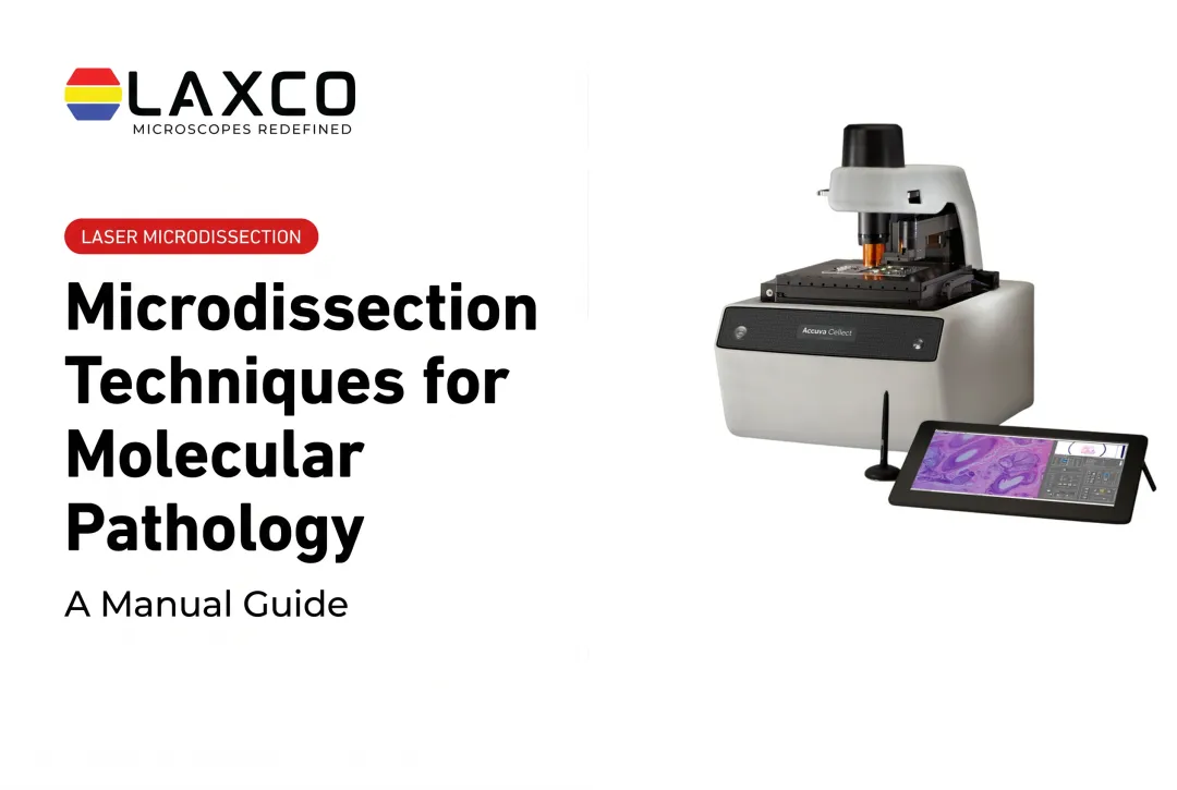
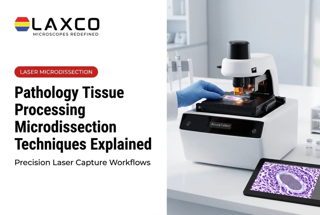
Laxco's Accuva Cellect Laser Capture Microdissection System isolates defined cell populations from tissue sections before extraction:
- Infrared (IR) laser: designed for capture of single cells or small numbers of cells.
- UV laser: designed for cutting dense structures and larger regions.
- Sample preparation: Laxco lists FFPE and fresh-frozen tissue, H&E-stained and LCM-optimized stains, immunofluorescence, unstained samples, and cell culture.
- Software workflow: a five-step workflow with visual quality control.
- Footprint: approximately 32% smaller than comparable microscope-and-laser systems, according to Laxco. Confirm the exact dimensions of the selected configuration before planning laboratory space.
Laxco lists training and 48-hour response times as part of its Accuva Cellect support offering. Accuva Cellect prepares selected tissue regions for downstream molecular analysis; it does not perform mass spectrometry. Laxco's Cellect Grant Assistance Program provides Grant Language Kits for NIH, NSF, HHMI, and DOD proposals involving LCM.
Frequently Asked Questions
Can archived FFPE tumor blocks be used for proteomics?
Yes. Archived FFPE tumor tissue can support mass-spectrometry-based proteomics when the extraction and digestion methods are validated for the samples. Heat-assisted retrieval and optimized chemistry can improve protein recovery, but they do not guarantee complete reversal of formalin-induced modifications. A 2025 study of matched rat-organ tissues demonstrated substantial proteome overlap under its tested conditions; performance in archived human tumors still depends on preservation history, tissue type, and workflow.
When is LCM worth the extra time compared with macrodissection?
LCM is worth considering when tumor cells are intermixed with stroma, immune cells, or necrosis, or when target regions are small. Macrodissection is faster and usually adequate for large, well-demarcated tumors with high tumor-cell content.
Which extraction steps does Laxco's LCM workflow cover?
Laxco's microgenomics workflow for Accuva Cellect lists fresh, frozen, and FFPE extraction, protein extraction, and mass spectrometry as downstream options. Laxco supplies the instrument and workflow components; the extraction chemistry and mass spectrometry are performed in your laboratory or core facility.
Which stains can I use before capturing tumor regions?
Laxco lists H&E staining, LCM-optimized stains, immunofluorescence, and unstained sections among the preparations supported by Accuva Cellect. For proteomics, choose a staining protocol that reliably identifies the target cells or tissue regions and is compatible with the intended protein-extraction and digestion workflow.
Staining effects depend on tissue preservation, staining chemistry, processing conditions, and the downstream analytical method. A 2025 review of mass-spectrometry-based FFPE proteomics discusses how pre-analytical variables, fixation-related modifications, and extraction methods affect protein recovery and analytical performance.
Researchers should validate the selected staining and extraction workflow with representative samples rather than assume that results reported for one preparation or instrument apply to Accuva Cellect.
What support does Laxco provide after an Accuva Cellect installation?
Laxco lists training and 48-hour response times for Accuva Cellect. Ask a Laxco specialist to confirm the training arrangements, service terms, and response coverage included with the selected configuration.