
Key Takeaways
- Fluorescence microscopy detects emitted light from fluorophores, which provides target-specific contrast.
- Widefield, confocal, multiphoton, and TIRF systems suit different sample thicknesses and imaging goals.
- Photobleaching, phototoxicity, and autofluorescence affect image quality and live-cell viability.
- System selection depends on sample type, fluorophores, resolution, speed, and analysis requirements.
How Fluorescence Imaging Works
A fluorophore absorbs light at one wavelength and emits light at a longer wavelength. Part of the absorbed energy is lost before emission, and the resulting wavelength difference is called the Stokes shift. Filters use this difference to separate emitted light from excitation light.
The Optical Path
A typical fluorescence microscope has six key elements:
- Light source: LED, arc lamp, or laser.
- Excitation filter: selects the wavelengths that excite the fluorophore.
- Dichroic mirror: reflects excitation light toward the sample and transmits emitted light.
- Objective: focuses excitation light and collects emitted light.
- Emission filter: blocks residual excitation light.
- Detector or camera: records the image.

Fluorophore Types
- Fluorescent proteins, such as GFP and its derivatives, can be expressed as genetically encoded reporters, fused to proteins of interest to study localization and dynamics, or incorporated into fluorescent biosensors.
- Organic dyes include DAPI for DNA and nuclear staining, as well as dyes designed to label specific organelles, membranes, or other cellular structures.
- Antibody conjugates detect specific targets by immunofluorescence.
Common Challenges
- Photobleaching: irreversible loss of fluorescence under prolonged or intense excitation.
- Phototoxicity: light-induced damage to live cells, which can occur before fluorescence loss is visible.
- Autofluorescence: native fluorescence from the sample that can obscure specific signal.
Illumination intensity, wavelength, and exposure time influence photobleaching and phototoxicity, so acquisition conditions should be optimized for the fluorophores and sample. For multichannel fluorescence observation, Laxco's SLi3Pro and SLi6Pro use patented SeBaLIT™ technology for real-time multichannel fluorescence imaging.
Types of Fluorescence Microscopes for Cell Imaging
Widefield (Epifluorescence) Microscopy
Widefield systems illuminate the entire field of view at once. They offer fast full-field acquisition and a large field of view. Because they do not reject out-of-focus light, image contrast decreases in thicker samples.
Laxco's inverted fluorescence systems relevant to conventional cell fluorescence imaging include:
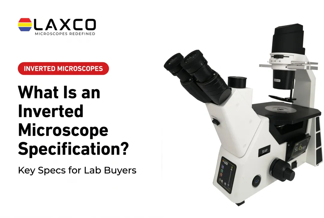
- SLi3Pro: supports common DAPI, FITC, and TRITC fluorochromes and is field-upgradeable to fluorescence and digital imaging.
- SLi6Pro: covers 390–662 nm and supports more than 110 fluorochromes, including CFP, YFP, and RFP.
Confocal Microscopy
Confocal systems use a pinhole to reject out-of-focus light, which provides optical sectioning and supports 3D reconstruction. Point-scanning confocal systems offer high resolution at slower acquisition speeds. Spinning-disk systems acquire faster, with some trade-offs in sectioning (PMC review). Confocal systems generally require more training than widefield systems.
Multiphoton and TIRF Microscopy
Multiphoton microscopy uses nonlinear excitation to confine fluorescence to the focal volume. It supports deeper imaging in scattering tissue. A review reported typical imaging depths of around 500 µm, depending on tissue scattering (PMC).
TIRF microscopy restricts excitation to a thin evanescent field near the coverslip, typically under about 100 nm deep. It is used for membrane-level and single-molecule studies.
| System type | Commonly used for | Acquisition speed | Relative complexity |
|---|---|---|---|
| Widefield | Thin samples, fast imaging | Fast | Lowest |
| Confocal | Optical sections, 3D imaging | Slower for point scanning | Higher |
| Multiphoton | Deep tissue and live samples | Depends on configuration | Highest |
| TIRF | Membrane and surface events | Fast | Specialized |

Applications of Cell Fluorescence Imaging
- Organelle visualization: organelle-specific dyes label mitochondria, nuclei, and cytoskeletal networks. Potentiometric dyes such as TMRM report changes in mitochondrial membrane potential.
- Protein interaction studies: FRET measures proximity between donor and acceptor fluorophores over roughly 10–100 Å. It is used to study protein-protein interactions and calcium signaling in living cells (PMC).
- Live-cell time-lapse imaging: tracking division, migration, and treatment responses requires balancing signal quality against light exposure.
- Cancer research, drug discovery, and developmental biology: fluorescent probes are used to follow cell-cycle progression, drug responses, and embryonic development.
- Immunofluorescence and FISH: labeled antibodies and nucleic-acid probes detect specific proteins and sequences, including chromosomal rearrangements.
For time-lapse imaging inside a CO₂ incubator, Laxco's CellLive CL22 supports brightfield, phase contrast, multichannel fluorescence, and Z-stack scanning with scheduled acquisition.
Image Analysis
Manual analysis of large image sets is time-consuming and can vary between analysts. Laxco's LEAP AI combines Accuva LEAP optics with Media Cybernetics Image-Pro AI. It follows an Image → Process → Measure → Report workflow with these capabilities:
- Pre-trained deep-learning models for AI-assisted segmentation.
- Batch processing and protocol-driven workflows, including batch-ready analysis protocols.
- Measurement and reporting tools.
- Optional AutoQuant deconvolution and 3D visualization and analysis.
Automated segmentation can reduce manual workload and some sources of analyst-to-analyst variability when validated for the dataset. It does not address photobleaching or phototoxicity, which depend on acquisition conditions.
Advantages and Limitations
Advantages
- High specificity through targeted labeling.
- Multiplexing of several fluorophores in one sample.
- Quantitative analysis of fluorescence intensity and localization when labeling, acquisition settings, and relevant calibration or normalization procedures are appropriately controlled.
- Compatibility with live and fixed samples.
Limitations
- Photobleaching limits total imaging time per sample.
- Phototoxicity can affect live cells during long sessions.
- Widefield and confocal systems have limited penetration in thick tissue.
- Advanced systems require more training and maintenance.
Choosing a Fluorescence Microscope
Define the following before comparing systems:
- Sample type and thickness: live cells, fixed monolayers, and thick tissue have different requirements.
- Fluorophore panel: match the excitation and emission ranges of all labels to available illumination and filters.
- Optical sectioning and resolution: determine whether the experiment requires rejection of out-of-focus fluorescence or 3D optical sections, and separately consider the spatial resolution needed for the structures of interest.
- Imaging speed: time-lapse work requires faster acquisition than static imaging.
- Analysis workflow: consider how images will be segmented and measured.
- Total cost of ownership: include training, service, and software as well as the instrument.
Frequently Asked Questions
Which Laxco microscope suits standard three-color fluorescence?
The SLi3Pro supports common DAPI, FITC, and TRITC fluorochromes using SeBaLIT™ illumination. It is field-upgradeable to fluorescence and digital imaging.
Which Laxco microscope supports a broader range of fluorophores?
The SLi6Pro extends spectral compatibility from approximately 390–662 nm and supports more than 110 fluorochromes, including options across cyan, yellow, and far-red ranges. It remains a 3-color multiplex fluorescence platform, so fluorochrome compatibility should not be interpreted as the number of labels that can be imaged simultaneously. Share your fluorophore requirements with a Laxco specialist to confirm the appropriate module and filter configuration.
Can I image live cells without removing them from the incubator?
Laxco's CellLive systems are designed for continuous imaging inside CO₂ incubators. The CellLive CL22 adds phase contrast, multichannel fluorescence, and Z-stack scanning to brightfield imaging.
When is confocal microscopy necessary instead of widefield?
Confocal microscopy is useful when out-of-focus light degrades images of thicker samples or when 3D reconstruction is required. For thin monolayers, widefield imaging is often sufficient and faster.