
Key Takeaways
- LCM isolates selected cells or regions from tissue sections under microscopic guidance, without tissue dissociation.
- RNA quality depends on tissue handling, staining conditions, and the time between staining and capture.
- Low-input library preparation methods are typically needed because LCM yields small amounts of RNA.
- Fresh-frozen tissue usually yields higher RNA integrity, while FFPE tissue offers access to archived samples.
- Published studies have used LCM-RNA-seq to profile distinct compartments in colorectal cancer and glioblastoma.
What Is Laser Capture Microdissection?
LCM is a microscopy-guided method for isolating selected cells or tissue regions. The operator views a stained section, marks the target, and uses a laser to capture or cut it. The captured material is then processed for DNA, RNA, or protein analysis.
LCM differs from other isolation methods in several ways:
- Manual microdissection uses needles or blades under a microscope. It depends on operator skill and is less suited to small or irregular targets.
- FACS and droplet methods sort or partition cells from a dissociated suspension. They do not retain the location of each cell in the tissue.
- LCM collects material directly from sections or cultures, so the histological context of each sample can be documented.
LCM throughput is lower than flow-based or droplet methods. It is most useful when the selection itself depends on morphology or location.
How LCM Works for RNA Studies
A typical workflow for RNA analysis follows these steps:
- Section tissue onto slides suited to the LCM system and the capture method.
- Stain the section with a protocol compatible with RNA preservation, such as a rapid hematoxylin and eosin (H&E) stain.
- Identify target regions under the microscope.
- Capture the selected cells or regions using the laser.
- Collect the material into an RNase-free tube or cap for extraction.

Time and RNA Integrity
RNA can degrade during staining and microdissection because residual water allows endogenous RNase activity. A 2008 study tested two approaches to reduce this: performing LCM under an argon atmosphere and using ethanol-based staining solutions (Clément-Ziza et al., RNA, 2008). Both methods stabilized RNA during microdissection for up to 90 minutes in that study.
General practices that help preserve RNA include:
- Cleaning surfaces and instruments with RNase-decontamination reagents
- Using nuclease-free reagents and RNase-free consumables
- Limiting freeze-thaw cycles of tissue blocks
- Keeping the time between staining and capture as short as practical
From Captured Cells to RNA-Seq Data
RNA Quality Assessment
The RNA Integrity Number (RIN) is a widely used measure of RNA integrity, scored from 1 (highly degraded) to 10 (highly intact), as described by Schroeder et al. (2006). RNA quality in LCM samples can be affected by tissue preservation, fixation, staining, handling conditions, and processing time. Small RNA input does not inherently indicate degradation, although very low quantities can make RNA-quality measurements less reliable or fall below an instrument's validated measurement range. For fragmented FFPE-derived RNA, DV200 is often a more useful metric than RIN when assessing library-preparation suitability.
Low-Input Library Preparation
Standard RNA-seq library kits assume more input than LCM usually provides. Low-input and amplification-based chemistries are therefore commonly used. Input requirements vary by kit, so they should be confirmed before planning the number of cells to capture.
Quality Control After Sequencing
Useful checks for LCM-RNA-seq data include:
- Gene-body coverage: degraded samples often show 3′ bias.
- Duplication rate: typically higher in low-input libraries.
- GC content distribution: can reveal amplification bias.
- Saturation analysis: shows whether sequencing depth is sufficient for low-abundance transcripts.
Results are commonly confirmed by qPCR on microdissected material and by immunohistochemistry on adjacent sections.

Fresh-Frozen and FFPE Tissue
The choice often depends on which samples are available.
| Factor | Fresh-frozen | FFPE |
|---|---|---|
| RNA integrity | Generally higher | Generally lower and fragmented |
| Availability | Requires frozen storage, usually collected prospectively | Widely available in pathology archives |
| Morphology | Adequate for many applications | Often well preserved |
| Typical use | Prospective studies needing higher RNA quality | Retrospective and archival cohorts |
A 2023 study compared whole-transcriptome sequencing of paired fresh, frozen, and FFPE cardiac tissue (PLOS ONE, 2023). RNA from FFPE tissue was highly degraded and yielded more intronic and intergenic reads. Protein-coding transcripts still showed high correlation across storage conditions (ρ > 0.94) in that study.
Published Applications
Matched Primary and Metastatic Colorectal Tumors
A 2020 protocol study used LCM to isolate pure cell populations from matched primary colorectal tumors and metastases (Scientific Reports, 2020). The authors reported an optimized protocol that reproducibly delivered intact RNA for RNA-seq and qPCR.
Histological Compartments in Glioblastoma
A study of archived FFPE glioblastoma tissue used LCM and RNA-seq to profile six histological contexts (Frontiers in Oncology). The regions were:
- Tumor core astrocytes
- Pseudopalisading astrocytes
- Perineuronal astrocytes in satellitosis
- Neurons with satellitosis
- Tumor blood vessels
- Normal blood vessels
The authors developed a customized protocol for RNA amplification and library construction from each microdissected portion.
Laxco Accuva Cellect for LCM-RNA-Seq Workflows
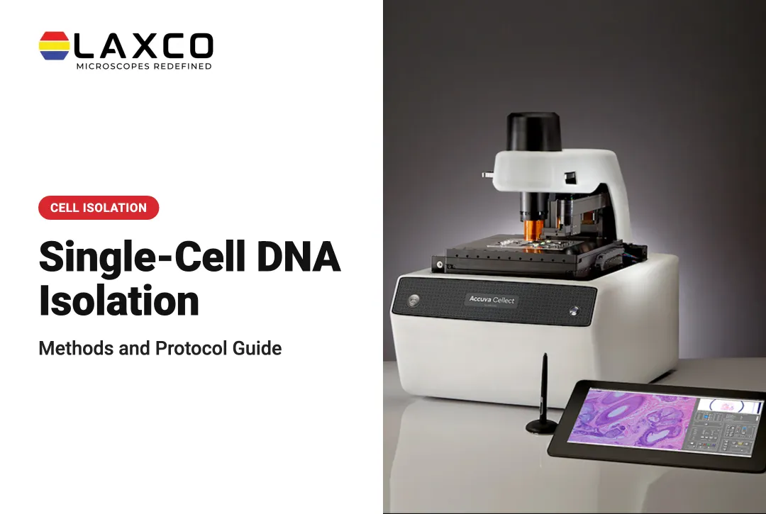
Researchers who need region-specific RNA from tissue sections may consider LCM. Laxco's Accuva Cellect Laser Capture Microdissection System is designed for this step:
- Dual-laser design: an infrared (IR) laser captures single cells or small numbers of cells. A UV laser cuts dense structures such as bone or plant tissue.
- Sample preparation: Laxco lists FFPE and fresh-frozen tissue, H&E-stained and LCM-optimized stains, immunofluorescence, unstained samples, and cell culture.
- Software workflow: a five-step workflow supports target selection, visual quality control, and documentation.
- Microgenomics workflow: Accuva Cellect supports the identification, selection, and collection of target cells or tissue regions. Captured material can then undergo DNA or RNA purification, amplification, and analysis through separate downstream laboratory workflows. CapSure LCM caps and compatible extraction methods are relevant to sample collection and processing.
- Footprint: approximately 32% smaller than comparable microscope-and-laser systems, according to Laxco. Confirm the exact instrument dimensions for the selected configuration.
Laxco offers the Accuva Cellect Laser Capture Microdissection System and lists training and 48-hour response times as part of its support offering. RNA extraction, library preparation, and sequencing are downstream laboratory or core-facility workflows, not analytical functions performed by the microdissection instrument.
Frequently Asked Questions
Which tissue preparations does Accuva Cellect support for RNA-seq?
Laxco lists FFPE and fresh-frozen tissue, H&E-stained and LCM-optimized stained sections, immunofluorescence, unstained samples, and cell culture. For RNA-seq, fresh-frozen tissue usually gives higher integrity, while FFPE gives access to archived cohorts.
How many cells should I capture for RNA-seq?
The required input depends on the cell type, RNA yield and integrity, and the library-preparation method. Choose a low-input RNA-seq workflow that is compatible with RNA extracted from microdissected material. Do not assume that every single-cell RNA-seq kit is suitable for extracted LCM RNA, because some workflows require intact cells or nuclei as input. Confirm the library kit's input requirements and run pilot captures to assess library complexity before using valuable tissue.
How do I protect RNA during microdissection?
Use RNase-controlled reagents, consumables, and handling practices, and minimize unnecessary delays between staining, microdissection, and sample stabilization. Follow the dehydration and drying requirements validated for the tissue, staining protocol, and LCM instrument; IR thermoplastic-film capture often requires appropriately dry sections, but full dehydration is not a universal rule for every UV-based workflow. A 2008 study by Clément-Ziza and colleagues reported improved RNA stability for up to 90 minutes under its tested argon-atmosphere or ethanol-based staining conditions. That finding should not be treated as a universal processing-time allowance. Proceed promptly to the lysis, stabilization, or storage conditions required by the downstream RNA workflow.
Can I see which region each RNA-seq sample came from?
Yes. Accuva Cellect uses a five-step software workflow with visual quality control, so selected regions can be documented before and after capture. This record links each library to the histology it represents.
What support is available when setting up LCM-RNA-seq?
Laxco lists training and 48-hour response times for Accuva Cellect. Confirm the specific training arrangements, service terms, and application assistance available for your planned tissue and RNA workflow. RNA extraction, library preparation, and sequencing remain separate downstream laboratory or core-facility procedures.