LCM is a microscopy-guided technique for isolating selected cells or tissue regions while retaining information about where those cells originated. Researchers use it to enrich target populations before DNA, RNA, or protein analysis, particularly when surrounding tissue could dilute the molecular signal.
The slide-by-slide outline below can be adapted for a laboratory presentation, training session, or research-methods lecture. The accompanying sections provide the technical explanations and references needed to develop each slide.
Key Takeaways
- LCM isolates targets from mixed tissue under direct microscopic visualization.
- Infrared (IR) lasers are used for film capture, and ultraviolet (UV) lasers for cutting.
- Manual macrodissection is efficient for large tissue regions, manual microdissection uses fine tools under magnification for smaller targets, and LCM enables more precise isolation of selected cells or closely intermixed populations.
- Captured material supports DNA, RNA, and protein analysis in oncology, neuroscience, and developmental biology research.
Laser Capture Microdissection PPT: 10-Slide Presentation Outline
Use this outline to prepare an introductory presentation for laboratory researchers, graduate students, or core-facility users. Each slide identifies the concepts to explain rather than prescribing a particular visual design. The supporting sections below provide additional technical context.
Slide 1 — What Is Laser Capture Microdissection?
- LCM isolates selected cells or tissue regions under microscopic guidance.
- The collected material can undergo downstream DNA, RNA, or protein analysis.
- The primary advantage is more selective sampling from heterogeneous tissue.
Presentation note: Explain the difference between isolating a target cell population and analyzing an entire tissue section.
Slide 2 — Why Tissue Heterogeneity Matters
- Tumors and other tissues contain multiple intermixed cell populations.
- Bulk extraction combines molecular signals from target and non-target cells.
- Selective collection can enrich the population relevant to the research question.
Presentation note: Use tumor epithelium surrounded by stromal and immune cells as an illustrative example.
Slide 3 — Main Components of an LCM System
- A microscope displays tissue morphology and candidate targets.
- A laser isolates selected cells or tissue regions.
- Software supports target selection and, depending on the system, capture documentation and quality control.
Presentation note: Distinguish the physical isolation step from downstream molecular analysis.
Slide 4 — Infrared Versus Ultraviolet Lasers
- Infrared film-capture systems use localized heating to attach selected cells to a thermoplastic collection surface.
- Ultraviolet systems cut around defined regions by laser ablation.
- Laser choice depends on target size, tissue properties, and collection requirements.
Presentation note: IR capture and UV cutting are different mechanisms. Do not describe all LCM instruments as using both.
Slide 5 — Tissue and Slide Preparation
- FFPE and fresh-frozen tissue may require different sectioning and preservation workflows.
- H&E staining, optimized staining, immunofluorescence, or unstained sections may assist target selection.
- Staining chemistry, dehydration, handling time, and section quality can affect downstream analytes.
Presentation note: There is no universal preparation protocol for every tissue and molecular assay.
Slide 6 — The LCM Workflow
- Prepare and mount the tissue section.
- Identify the target under the microscope.
- Select the region for capture or cutting.
- Isolate and collect the material.
- Inspect the result and proceed to a validated extraction workflow.
Presentation note: Confirm that target selection and capture have occurred before interpreting downstream data.
Slide 7 — LCM Versus Manual Dissection
- Manual macrodissection efficiently collects relatively large, well-defined regions.
- Manual microdissection uses fine tools under magnification for more selective tissue removal.
- LCM offers instrument-guided isolation for targets that are difficult to separate manually, including selected individual cells.
Presentation note: LCM is not automatically necessary for every specimen. Match the method to the required purity and resolution.
Slide 8 — Research Applications
- Oncology: enrichment of tumor and microenvironment compartments.
- Neuroscience: isolation of defined structures or neuronal populations.
- Developmental biology: collection of spatially defined tissue regions.
- Spatial molecular research: targeted collection before RNA sequencing or proteomic analysis.
Presentation note: LCM preserves the identity and location of collected material; it does not independently perform sequencing or mass spectrometry.
Slide 9 — Limitations and Experimental Controls
- Low-input samples can challenge nucleic-acid or protein recovery.
- Staining, processing time, tissue quality, and collection settings influence results.
- Capture purity and downstream assay sensitivity require validation.
Presentation note: Document the sample source, selected regions, collection method, and relevant quality-control results.
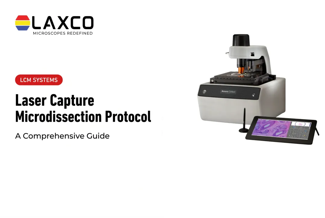
Slide 10 — Example System: LAXCO Accuva Cellect
- Accuva Cellect combines IR film capture and UV laser cutting in one LCM system.
- Its five-step software workflow supports selected-cell isolation, visual quality control, and documentation.
- The system supports research sample preparation across multiple tissue and staining formats.
- LAXCO offers application training, technical support, and grant-assistance resources.
Presentation note: Accuva Cellect performs the tissue-isolation step. DNA extraction, sequencing, and proteomics are separate downstream processes.
How to Use This Presentation Outline
For an introductory lecture, use the 10 slides in order. For a laboratory training session, expand Slides 5, 6, and 9 with locally validated sample-preparation procedures and quality-control examples. For instrument evaluation, expand Slide 10 to compare the laboratory's sample types, throughput requirements, and technical support needs with Accuva Cellect's documented capabilities.
This is a presentation outline, not a downloadable PPTX file.
What Is Laser Capture Microdissection?
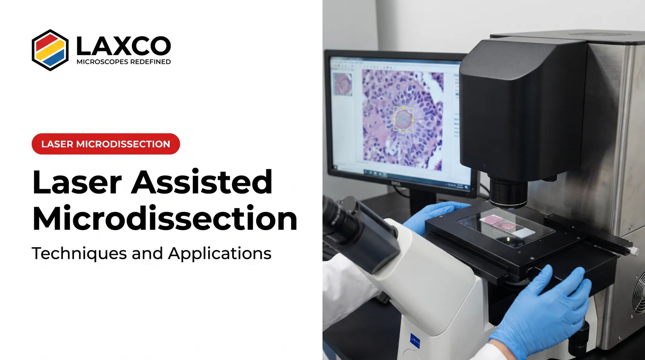
LCM separates selected cells or regions from surrounding tissue while the sample is viewed under a microscope. The operator selects the target in software, and the laser performs the capture or cut.
Origins
LCM was developed at the National Institutes of Health by Michael Emmert-Buck, Lance Liotta, and colleagues. Their 1996 paper in Science described capture using a thermoplastic film activated by an infrared laser (Science, 1996). The authors demonstrated downstream PCR and enzyme recovery from captured cells.
The Core Principle
An LCM system combines three components:
- A microscope for visualizing the section.
- A laser for capture or cutting.
- Software for selecting targets.
Common sample types include FFPE tissue, fresh-frozen sections, cytology preparations, and cultured cells.
Why purity matters: when non-target cells are included, DNA, RNA, and protein results reflect a mixture. This can mask a real signal or create a misleading one.
How LCM Works
A typical LCM workflow follows five steps:
- Section the tissue and mount it on a slide.
- Visualize the sample under the microscope.
- Select target cells in the software.
- Capture or cut the target with the laser.
- Collect the material for extraction.

Laxco's Accuva Cellect software uses a five-step workflow with visual inspection for quality control and documentation.
Two Laser Types
| Laser type | Mechanism | Commonly used for |
|---|---|---|
| Infrared (IR) | Locally melts a thermoplastic film, which adheres to and lifts the selected cells | Single cells or small numbers of cells |
| Ultraviolet (UV) | Photoablates tissue along a defined path to cut out a region | Larger regions and dense structures |
A 2013 protocol review describes both mechanisms (PMC, 2013). Cutting width and capture size depend on tissue type, stain, section thickness, and instrument settings.
Collection Methods
- Film capture on a cap: cells adhere to a thermoplastic film on a collection cap and are lifted from the slide.
- Gravity-assisted collection: cut material falls into a collection tube below the slide.
- Laser pressure catapulting: a laser pulse propels the cut material into a collector above the slide.
Each approach has different requirements for sample preparation and consumables. Protocol time depends on sample complexity, staining, and the number of regions collected.
Downstream Applications
Captured material can be used for:
- DNA genotyping and loss-of-heterozygosity analysis.
- RNA profiling, including qPCR and RNA-seq.
- cDNA library generation.
- Proteomics by mass spectrometry.
Manual Microdissection Compared with LCM
Manual microdissection uses needles, scalpels, or micromanipulators to remove tissue regions by hand under a microscope. A 2022 narrative review noted that manual dissection is generally cheaper and faster than LCM (Journal of Xiangya Medicine, 2022). Manual macrodissection generally suits larger, clearly defined regions, while manual microdissection can isolate smaller, visually identifiable targets under magnification when they are accessible with fine tools.
Manual methods have limitations:
- Operator variability: results depend on individual technique.
- Contamination: scraping can include non-target cells.
- Resolution: isolating single cells or small structures by hand is difficult.
LCM also has trade-offs. The same review notes that laser heat, collection time, and nuclease or protease activity can affect analyte quality. Protocol optimization addresses these factors.
Manual macrodissection is often sufficient for larger, clearly defined tissue regions. Manual microdissection provides finer selection when targets are visible and accessible under magnification. LCM becomes more useful when individual cells, rare populations, or closely intermixed structures require higher-precision, laser-guided isolation.
Research Applications
Oncology
A common use of LCM is separating tumor cells from surrounding stroma. Scraping whole sections dilutes tumor DNA and RNA with non-neoplastic material, which reduces assay sensitivity.
Laser microdissection can enrich tumor epithelium or other histologically defined compartments before molecular analysis. Separating selected tumor-rich regions from surrounding stromal and immune-cell populations helps researchers investigate molecular signals associated with the target tissue rather than the entire specimen.
Depending on the experimental design, collected material can be used for DNA analysis, RNA profiling, or protein extraction followed by a separately performed proteomics workflow. The choice of downstream assay determines the preservation, input, and quality-control requirements.
Drug Discovery
LCM can isolate specific cell populations from treated tissue to measure cell-type-specific responses and biomarkers.
Developmental Biology
LCM combined with RNA-seq can sample small numbers of cells from defined embryonic structures. This tracks expression changes as tissues form.
Neuroscience
LCM can isolate defined structures, such as amyloid plaques or specific neuronal layers, for proteomic or transcriptomic comparison with surrounding tissue.
Spatial Biology
LCM is increasingly used as a sampling step in spatial proteomics workflows. Image-based selection of cell populations is followed by microdissection and mass spectrometry.
Choosing an LCM System
Consider the following before selecting a system:
- Laser type: IR for film capture of small numbers of cells, UV for cutting larger or dense regions, or both.
- Sample compatibility: FFPE, fresh-frozen, cytology, and cell culture support.
- Collection method: cap-based film capture, gravity collection, or catapulting.
- Software: how targets are selected, documented, and checked for quality.
- Support: training, application guidance, and service response times.
Laxco's Accuva Cellect Laser Capture Microdissection System provides:
- An IR laser designed for film capture of single cells or small numbers of cells.
- A UV laser designed for cutting dense structures, such as bone or plant tissue.
- A five-step software workflow with visual quality control and documentation.
- Documented support for FFPE and fresh-frozen tissue, H&E-stained and LCM-optimized stains, immunofluorescence, unstained samples, and cell culture.
- CapSure LCM caps as part of the capture workflow.
- On-site and virtual applications training, live technical support, and a 48-hour response time.
The Cellect Grant Assistance Program provides Grant Language Kits for NIH, NSF, HHMI, and DOD proposals that include LCM.
Frequently Asked Questions
What is the difference between IR and UV laser microdissection?
IR lasers melt a thermoplastic film that adheres to selected cells, which suits single cells or small groups. UV lasers cut along a defined path, which suits larger regions and dense tissue. Accuva Cellect includes both lasers.
What samples can be used with Accuva Cellect?
Laxco lists FFPE and fresh-frozen tissue, H&E-stained and LCM-optimized stained sections, immunofluorescence, unstained samples, and cell culture. Confirm compatibility with your downstream assay during planning.
Does LCM affect molecular analysis?
With an optimized protocol, LCM-captured material supports DNA, RNA, and protein analysis. Staining, dehydration, laser settings, and collection time can affect analyte quality, so pilot runs are advisable.
When is manual microdissection sufficient?
Manual microdissection is appropriate when the target can be clearly identified under magnification and separated using a fine tool without unacceptable contamination. For larger, well-defined regions, manual macrodissection may be sufficient and more efficient. LCM is better suited to selected individual cells, rare or scattered populations, and closely intermixed structures requiring finer isolation.