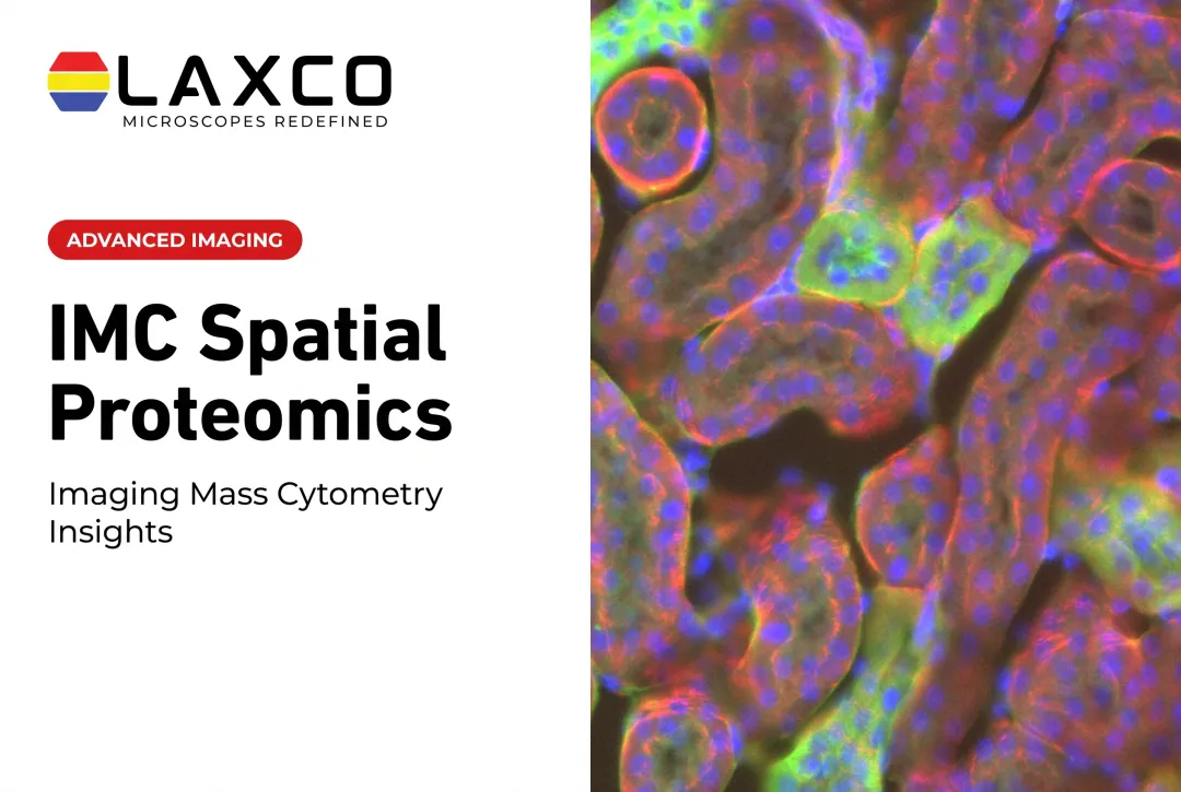
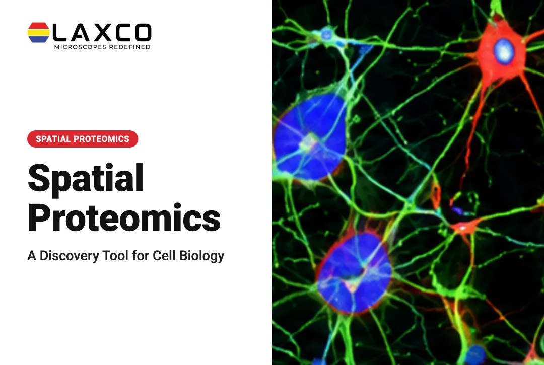
Conventional immunofluorescence is generally constrained by the number of fluorophores that can be distinguished in one imaging pass. Hyperplex methods extend multiplexing through repeated fluorescence imaging cycles, DNA-barcoded antibody readouts, or metal-tagged antibody detection. This guide explains the principal technique families, applications, panel-design considerations, and factors to evaluate when selecting an approach.
Key Takeaways
- Hyperplex methods measure many proteins on one section while retaining tissue architecture.
- The three main families are cyclic fluorescence, DNA-barcode readout, and metal-tag mass cytometry imaging.
- Applications include tumor microenvironment mapping, immunotherapy research, and neural tissue studies.
- Antibody validation and image analysis capacity affect data quality more than the maximum plex number.
- Discovery studies often use large panels, followed by smaller validated panels in larger cohorts.
What Is Hyperplex Spatial Proteomics?
Hyperplex spatial proteomics commonly uses labeled antibodies to measure multiple proteins in tissue sections without homogenizing the entire specimen. Unlike conventional bulk proteomics, spatial methods retain information about where signals originate within the tissue.
Imaging-based methods can generate spatially resolved protein-marker images. Cell segmentation and downstream analysis may then associate marker signals with individual cells and their neighbors. Other spatial protein-profiling methods instead generate measurements linked to defined regions of interest rather than independent measurements for every cell.
Nature Methods named spatial proteomics its Method of the Year for 2024 (Nature Methods, 2024).
Several fluorescence-based hyperplex methods use repeated imaging cycles, but the preparation and signal-removal procedures differ between technologies.
In cyclic immunofluorescence, researchers may stain tissue with successive antibody sets and remove or inactivate fluorescence between imaging rounds. In DNA-barcoded approaches such as CODEX, tissue is generally stained with DNA-conjugated antibodies before repeated cycles of fluorescent reporter hybridization, imaging, and reporter removal.
A representative cyclic fluorescence workflow is:
Prepare and label the tissue. Apply a validated antibody panel using the labeling method required by the selected assay, such as fluorescent antibodies or DNA-barcoded antibody conjugates.
Generate fluorescence for the current cycle. Apply or activate the appropriate fluorescent labels or reporter oligonucleotides.
Acquire the fluorescence image. Record the selected channels using suitable microscope settings and document the tissue position.
Remove or inactivate the cycle's signal. Use the validated stripping, bleaching, or reporter-removal procedure and repeat the appropriate steps to image the remaining targets.
Register and analyze the images. Align the acquired image rounds into a multiplexed dataset, assess signal quality, and perform validated cell segmentation when individual-cell measurements are required.
Imaging mass cytometry and multiplexed ion beam imaging use destructive metal-isotope acquisition instead of this repeated fluorescence-imaging workflow.
Conventional immunofluorescence typically measures a few targets per section. Cyclic and barcode-based methods build larger panels across rounds instead of in one imaging pass.

Core Technique Families
Cyclic Fluorescence Methods
Cyclic immunofluorescence repeatedly applies suitable antibody staining, fluorescence imaging, and signal-removal or signal-inactivation steps to the same tissue section. Each round measures a limited number of channels, and measurements from successive rounds are registered to build a larger protein-marker dataset.
Some cyclic methods can use compatible conventional fluorescence microscopes or slide scanners, but a validated hyperplex workflow requires more than owning an imaging instrument. Laboratories must consider staining chemistry, signal removal, repeatable slide positioning, tissue preservation, image registration, and potentially additional automation or fluid-handling equipment.
Considerations: total workflow time generally increases as additional cycles are required. Repeated processing may affect tissue integrity or antigen detectability, while registration errors can misassign signals between cells. Assay-specific validation and suitable quality controls are essential.
DNA-Barcode Methods
DNA-barcoded spatial protein methods use antibodies linked to distinct oligonucleotide sequences, but the readout and spatial resolution vary between platforms.
In fluorescence-imaging approaches such as CODEX, antibody-associated DNA barcodes are identified through repeated hybridization of complementary fluorescent reporters. Signals can then be registered and analyzed to produce cell-associated measurements when imaging resolution and segmentation are suitable.
Other barcode-based spatial profiling systems release or collect oligonucleotide tags from selected tissue regions for sequencing or digital counting. These methods may produce region-associated protein measurements rather than continuous single-cell protein images.
Considerations: the reported number of measurable proteins, imaging requirements, molecular readout, and spatial resolution are platform-dependent. Confirm whether a stated plex number refers to imaging individual cells or profiling larger tissue regions, and whether fluorescence imaging, sequencing, or another detection method is required.
Metal-Tag Mass Cytometry Imaging
Imaging mass cytometry and multiplexed ion beam imaging label antibodies with metal isotopes and measure them by mass spectrometry. Tissue autofluorescence does not affect these readouts.
| Factor | Cyclic fluorescence | Metal-tag spatial imaging (IMC or MIBI) |
|---|---|---|
| Background | Tissue autofluorescence and fluorescence-channel crosstalk may require correction | Tissue autofluorescence does not contribute to metal-isotope detection, but isotope-channel interference and other analytical artifacts still require control |
| Acquisition | Individual fluorescence imaging rounds may be relatively fast, but repeated staining and imaging can increase total workflow time | Acquisition speed depends on instrument, sampled area, and resolution; typically requires dedicated scanning or ablation equipment |
| Sample preservation | The same section may undergo repeated cycles, but processing can affect tissue integrity or antigen signal | IMC laser ablation or MIBI ion-beam sampling consumes the analyzed material |
| Equipment and workflow | Compatible fluorescence imaging plus validated staining, signal removal, registration, and analysis procedures | Dedicated metal-tag imaging instrumentation and appropriate image reconstruction and analysis |
Applications
Tumor Microenvironment Mapping
Staining for immune, stromal, and tumor markers on one section shows how cell types are arranged relative to one another. Studies have used multiplexed imaging to classify fibroblast and immune phenotypes and relate their spatial patterns to patient outcomes.
Immuno-Oncology
Spatial arrangement of immune cells, not only their numbers, has been associated with immunotherapy response in research studies. Multiplexed imaging supports investigation of these patterns as candidate biomarkers.
Neuroscience
Hyperplex panels can map cell neighborhoods in brain tissue, including immune and glial interactions in tumors and neurodegenerative disease.
From Discovery to Validation
A common, but not universal, study-design strategy is to move from exploratory profiling to independently validated findings:
- Discovery: broad protein-marker panels are used to investigate spatial patterns and generate candidate biological hypotheses.
- Validation: candidate patterns are evaluated using independent samples, reproducible analysis procedures, and appropriate orthogonal assays. A smaller targeted marker panel may be used when suitable.
- Translation: promising, reproducible findings can be evaluated in larger and more representative cohorts. Additional analytical and clinical validation is required before findings are considered for diagnostic or treatment-related use.
Panel size and cohort size are research-design decisions rather than mandatory properties of each stage.
Panel Design and Workflow Considerations
Antibody Validation
Antibody clone selection, titration, and channel or isotope assignment determine whether results reflect biology or artifacts. Each batch should include:
- Single-stain titrations
- Positive and negative control tissues
- Co-localization checks for markers expected to overlap or remain separate
Tissue Preparation and Region Selection
Tissue preservation, staining, and region-of-interest selection affect the quality and interpretation of hyperplex measurements. When additional molecular profiling is needed, laser capture microdissection (LCM) can isolate selected cells or tissue regions for separate sequencing or mass-spectrometry analysis.
However, compatibility depends on when and how imaging is performed. IMC laser ablation and MIBI ion-beam sampling consume the analyzed tissue, so the same sampled material cannot subsequently be recovered by LCM. Complementary analyses may instead use aligned serial sections or a validated imaging-before-capture workflow.
LCM preserves a record of the captured region's origin, but pooled capture produces a combined molecular profile rather than a continuous spatial protein map. RNA extraction, sequencing, and mass spectrometry are downstream laboratory procedures performed separately from the capture instrument.
Image Analysis
Segmenting cells across many channels requires automated tools. Deep-learning segmentation can convert multichannel images into cell-level measurements, but results should be checked against manually annotated images.
Choosing an Approach
Consider five factors:
- Plex level: the number of targets needed.
- Spatial resolution: cellular or subcellular detail.
- Tissue compatibility: FFPE, fresh-frozen, and tissue type.
- Throughput: samples and tissue area per run.
- Analysis infrastructure: data storage, computing, and bioinformatics support.
Costs include instruments, antibody panels, validation time, and analysis. Laboratories without in-house platforms can use core facilities for specific projects.
Laxco Tools for Spatial Proteomics Workflows
SLi6Pro: an inverted widefield fluorescence microscope using SeBaLIT™ illumination across approximately 390–662 nm. It supports three-color multiplex fluorescence imaging and compatibility with more than 110 fluorochromes. This dye-compatibility figure does not represent simultaneous imaging of 110 markers. SLi6Pro is not documented as a validated, standalone hyperplex spatial proteomics system. Any cyclic hyperplex workflow would require additional compatible staining, acquisition, registration, and analysis procedures.
Accuva Cellect: a laser capture microdissection system combining an IR laser designed for individual cells and small cell populations with a UV laser intended for cutting dense structures. Laxco lists FFPE, fresh-frozen, and immunofluorescence-prepared samples among its supported preparations. Cellect can support complementary region isolation, but it does not perform hyperplex imaging, RNA sequencing, or mass spectrometry.
LEAP AI: a separate research microscopy and image-analysis platform combining Accuva LEAP optics with Media Cybernetics Image-Pro AI. It supports AI-assisted image segmentation, measurement, batch processing, and reporting. It is not documented as a dedicated high-plex spatial proteomics pipeline or an automatic controller for Cellect laser capture. Compatibility with externally generated multiplexed image datasets should be confirmed rather than assumed.
Laxco provides research instrumentation and lists training and 48-hour response times for Accuva Cellect. Confirm available support arrangements for each product separately. Laxco does not provide hyperplex spatial proteomics as a laboratory analysis service.
Frequently Asked Questions
How is hyperplex imaging different from conventional immunofluorescence?
Conventional immunofluorescence generally measures a limited number of markers in a single imaging pass because fluorescent signals can overlap. Hyperplex spatial proteomics extends multiplexing through repeated fluorescence imaging cycles, DNA-barcoded antibody readouts, or metal-tagged antibody detection.
The spatial information depends on the platform. Imaging-based methods can support cell-level protein measurements when resolution and image segmentation are appropriate. Other methods generate measurements associated with tissue regions rather than independent measurements for every cell.
Can I run cyclic immunofluorescence on a Laxco microscope?
The SLi6Pro supports three-color multiplex fluorescence imaging across an expanded 390–662 nm range and is compatible with more than 110 fluorochromes. However, those capabilities alone do not establish compatibility with a complete cyclic hyperplex protocol.
Cyclic immunofluorescence additionally requires validated staining and signal-removal chemistry, repeatable acquisition, tissue stability across cycles, image registration, and appropriate analysis software. Confirm whether the intended protocol and any additional hardware or software are compatible with the selected Laxco configuration before planning a study.
How does LCM fit into a hyperplex study?
Hyperplex imaging can identify tissue regions or cell neighborhoods for complementary molecular investigation. LCM may isolate corresponding material from aligned serial sections or from the same section when the preceding staining and imaging workflow is compatible with later capture.
Regions already consumed by IMC laser ablation or MIBI ion-beam acquisition cannot be recovered by LCM. After capture, separate downstream sequencing or mass-spectrometry methods can provide additional molecular measurements. These results remain associated with the documented captured region and do not automatically generate individual-cell profiles or a continuous spatial map.
Can Accuva Cellect isolate regions identified by immunofluorescence?
Yes. Laxco lists immunofluorescence among the preparations supported by Accuva Cellect, along with FFPE and fresh-frozen tissue. Confirm that your staining and mounting steps are compatible with the downstream protein or nucleic acid extraction.
How can I standardize cell segmentation across many channels?
For supported research microscopy workflows, Laxco's LEAP AI combines Accuva LEAP optics with Media Cybernetics Image-Pro AI and provides AI-assisted segmentation, measurement, and batch processing.
However, LEAP AI is a separate microscopy and image-analysis platform, not a documented dedicated hyperplex spatial proteomics analysis pipeline. Compatibility with externally generated high-plex datasets, file formats, and channel configurations should be confirmed with Laxco rather than assumed.
Regardless of the software selected, validate cell segmentation against manually annotated images, assess performance across relevant tissue types and staining conditions, and use consistent quality-control procedures before processing a full cohort.