Key Takeaways
- Microdissection isolates targeted cells or regions from tissue sections for cleaner molecular analysis.
- The main approaches are macrodissection, manual microdissection, and laser capture microdissection (LCM).
- It is used in cancer genomics, biomarker research, and studies of heterogeneous tissue.
- Results depend on tissue quality, staining, operator skill, and equipment.
- Microdissection is not needed for homogeneous samples or when bulk analysis meets the study's purity requirements.
Where Microdissection Fits in Tissue Processing
Routine histology processing includes grossing, fixation, dehydration, clearing, infiltration, embedding, sectioning, and staining. Microdissection is an additional step after sectioning, used when molecular analysis requires a defined cell population.
Bulk analysis extracts material from the whole section. A tumor section may contain:
- Cancer cells
- Normal stroma
- Inflammatory infiltrate
- Necrotic tissue
- Blood vessels
Combining all of these can lower the proportion of tumor-derived DNA or RNA. If tumor content is low, mutations may fall below the detection limit of the assay. Enriching tumor cells before extraction increases tumor content in the sample. Tumor-content thresholds and enrichment practices for NGS are discussed in published AMP/CAP NGS guidelines.
How Microdissection Works

Inputs:
- A tissue section from fresh-frozen or formalin-fixed paraffin-embedded (FFPE) tissue
- A stain or reference slide for identifying the target
- Manual tools or a laser microdissection instrument
Step 1: Slide Preparation and Target Identification
The section is mounted, stained if needed (commonly with H&E or immunohistochemistry), and reviewed under a microscope. A pathologist or researcher marks the region of interest based on morphology or staining pattern.
Step 2: Isolation of the Target
- Macrodissection removes a large, marked tumor area from the slide, typically with a blade or scalpel, when the target region is large and well demarcated.
- Fine manual microdissection uses a needle, scalpel tip, or similar tool under magnification to collect smaller marked regions with greater operator control than bulk scraping.
- Laser capture microdissection (LCM) uses a laser under direct microscopic visualization to capture or cut selected cells or regions with cell-level precision.
Step 3: Collection and Processing
The isolated material is transferred to a tube or collection cap and processed for DNA, RNA, or protein extraction.
Applications
- Cancer genomics: enriching tumor cells for mutation and copy number analysis.
- Single-cell and small-population studies: isolating cells of one type from mixed tissue.
- Developmental biology: isolating defined embryonic regions for expression profiling.
- Biomarker research: comparing purified cell populations across tumor subtypes.
A 2019 study in Genome Research combined LCM with a low-input RNA sequencing method to profile individual cells and small groups from archival breast tissue (Foley et al., 2019).
Factors That Affect Results
- Tissue and slide quality: fixation and section thickness affect how cleanly cells separate.
- Equipment: manual tools cost less but are less precise for small targets. Laser systems are more precise but require training.
- Sample scale: microdissection adds time per sample, which matters in high-volume workflows.
- Purity limits: imprecise targeting can still collect surrounding cells, especially with manual methods.
When Microdissection May Not Be Needed
- The tissue is largely homogeneous.
- Tumor content is already high enough for the assay.
- The research question concerns the whole tissue rather than a specific population.
- Spatial methods that measure expression in intact sections are better suited to the question.
Macrodissection is faster and less expensive for large tumors with clear boundaries, when cell-level resolution is not required.

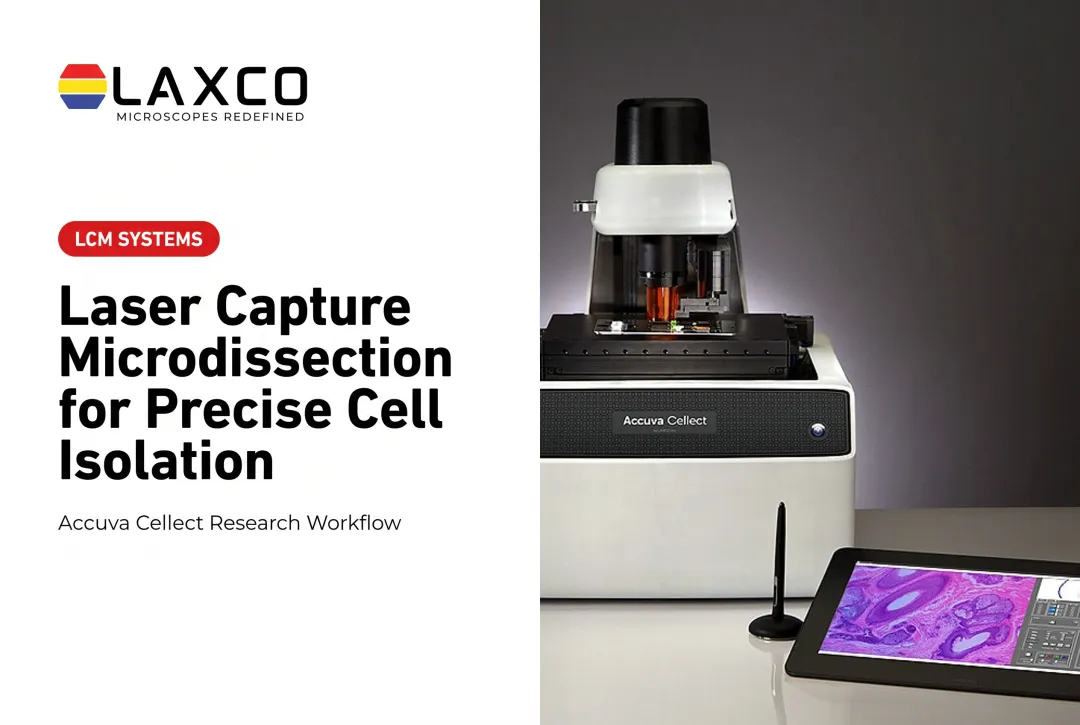
Laxco Accuva Cellect
Laxco's Accuva Cellect Laser Capture Microdissection System is a research instrument for isolating selected cells or regions in pathology-related research. It is distinct from a clinical diagnostic workflow. Documented features include:
- Infrared (IR) laser: designed for capture of single cells or small numbers of cells.
- UV laser: designed for cutting dense structures, such as bone or plant tissue.
- Sample preparation: Laxco lists FFPE and fresh-frozen tissue, H&E-stained and LCM-optimized stains, immunofluorescence, unstained samples, and cell culture.
- Collection: compatible with CapSure LCM caps.
- Software workflow: a five-step workflow with visual quality control before extraction.
Laxco lists training and 48-hour response times for Accuva Cellect. Confirm available training formats and service terms with Laxco. Laxco is not a commercial testing service and does not perform microdissection or molecular testing as a laboratory service. Its Cellect Grant Assistance Program offers Grant Language Kits for NIH, NSF, HHMI, and DOD proposals.
Frequently Asked Questions
When does a tumor sample need microdissection before testing?
Whether a tumor sample needs microdissection depends on the assay's validated tumor-content and purity criteria, not on a single universal threshold. When tumor cells are a small fraction of a heterogeneous section, enrichment can help keep mutations above the detection limit. When tumor content already meets the assay's requirements, microdissection is often unnecessary. Pathology practice guidance on specimen handling and molecular testing is summarized in published pathology guidelines.
Should I use macrodissection, manual microdissection, or LCM?
Choose by target size, required purity, throughput, and available equipment. Macrodissection suits large, well-defined tumor areas when cell-level resolution is not required. Fine manual microdissection suits smaller marked regions under magnification. LCM suits small, scattered, or intermixed targets where cell-level precision and documented selection matter.
How does fixation affect microdissected samples?
Formalin fixation preserves morphology but crosslinks and fragments nucleic acids and proteins, so molecular recovery from FFPE is typically lower and more variable than from fresh-frozen tissue. Fresh-frozen tissue generally gives better molecular quality when integrity is critical. Extraction methods should match the fixation used and the downstream assay; results depend on protocol and sample history rather than a universal FFPE-versus-frozen rule.
Which sample types does Accuva Cellect support?
Laxco lists FFPE and fresh-frozen tissue, H&E-stained and LCM-optimized stained sections, immunofluorescence, unstained samples, and cell culture for Accuva Cellect. Accuva Cellect is a research instrument. Its ETL mark is an electrical safety certification for North America and is not clinical diagnostic authorization or regulatory clearance.
How much bench space does Accuva Cellect need?
Accuva Cellect has a compact footprint, which Laxco describes as approximately 32% smaller than comparable microscope-and-laser systems. Confirm dimensions with Laxco when planning bench space.