Key Takeaways
- LCM isolates selected cells or regions from tissue sections while documenting their morphology and location.
- Infrared (IR) thermoplastic-film capture and ultraviolet (UV) cutting use different isolation and collection mechanisms. Both can isolate small targets, including individual cells, depending on the instrument and preparation.
- Fixation, staining, and slide preparation affect the quality and quantity of recovered material.
- Captured material is used for PCR, RNA sequencing, and proteomics.
- Image analysis software can help standardize target selection across operators.
What Is Laser Capture Microdissection?
LCM combines a microscope with a laser. The researcher identifies cells or a region on a stained or unstained section, and the laser captures or cuts the target for collection.
Manual microdissection compared with LCM:
- Manual microdissection uses blades or needles. It suits large target areas but is less precise for small targets, depends on operator dexterity, and more readily collects neighboring cells.
- LCM uses laser-assisted capture or cutting to isolate selected targets under microscopic guidance. Depending on the system, a laser activates a collection surface or cuts around the target. This can provide greater precision than manual dissection for small or closely intermixed cell populations.
A typical LCM workflow:
- View the section under the microscope.
- Mark the target region in the software.
- Capture or cut the target with the laser.
- Inspect the slide and collected material for quality control.
- Recover the collected material using the instrument's compatible collection cap, tube, or other vessel, and prepare it for the validated downstream extraction or analysis workflow.

How LCM Works
Infrared Capture
In IR capture, a thermoplastic film is positioned over the section. A low-energy infrared laser pulse melts the film onto the selected cells, which are lifted from the slide with the film. This approach suits single cells and small groups.
UV Cutting
In UV cutting, an ultraviolet laser cuts around the region of interest. The excised piece is then transferred to a collection device by gravity or another laser-assisted transfer method, depending on the instrument design. With suitable optics and workflow, UV cutting can isolate individual cells as well as larger regions. Suitability for dense tissue depends on the laser, optics, slide type, and validated protocol; UV cutting is commonly used for dense or tough tissues such as bone and plant tissue.
Some systems include both lasers so that the method can be matched to each target.
Magnification
Higher objective magnification allows precise targeting of individual cells. Lower magnification provides a wider field of view for selecting larger regions.
Image Analysis for Target Selection
Target selection has traditionally depended on operator judgment, which can vary between people and sessions. Deep-learning segmentation can identify candidate cells or regions consistently, while the researcher confirms the final targets.
Tissue Preparation
Fresh-Frozen and FFPE Tissue
- Fresh-frozen tissue generally preserves RNA better, but morphology can be less distinct.
- Formalin-fixed paraffin-embedded (FFPE) tissue preserves morphology and allows use of archived samples, but formalin crosslinking complicates nucleic acid extraction.
Adapted extraction protocols can improve RNA recovery from small microdissected FFPE samples (Amini et al., 2017).
Fixatives
- Precipitating fixatives such as ethanol and acetone generally preserve nucleic acids better.
- Crosslinking fixatives such as formalin preserve morphology but fragment and modify nucleic acids.
Staining and Drying
Targets must be visible before they can be selected. Common stains include:
- H&E for general morphology
- Toluidine blue or other quick stains for contrast
- Immunohistochemistry or immunofluorescence for marker-based identification
Slide preparation and dehydration requirements depend on tissue, stain, collection mechanism, and downstream assay. IR thermoplastic-film capture generally requires appropriately dry sections so the film can adhere to the selected targets. UV cutting workflows may use different drying or membrane-slide steps. Follow the instrument-validated protocol for the tissue type, stain, and collection method rather than applying one universal rule.
Slides
Membrane slides are commonly used for UV cutting, while glass slides are common for IR capture. Choose slides that suit the capture method and tissue type.
Research Applications
Cancer research: LCM enables researchers to isolate selected tumor regions separately from surrounding stroma, necrotic tissue, and immune-cell-rich compartments. The collected material can undergo downstream DNA, RNA, or protein analysis to investigate molecular differences between tissue regions. This targeted approach helps reduce signal mixing from unrelated cell populations, although the resulting measurements represent the collected cells or regions rather than automatically providing independent single-cell profiles.
Neuroscience and developmental biology: LCM isolates neighboring cell types or structures that look similar but differ in gene expression.
Drug discovery: LCM supports measurement of target expression in specific tissue compartments.
Single-cell and spatial biology: LCM can serve as an isolation step before omics analysis, while keeping a record of each sample's location.
Advantages and Limitations
Advantages:
- Precision down to single cells when needed
- Documented morphology and location of each sample
- Compatibility with PCR, sequencing, and proteomics
Limitations:
- Dedicated instruments and consumables add cost.
- Operators need training, and technique affects results.
- Small inputs often require amplification or highly sensitive downstream methods.
- Throughput is lower than bulk extraction.
Choosing an LCM System
- Laser options: IR, UV, or both.
- Magnification range: suitable for your typical target size.
- Sample compatibility: fixation types, stains, and slides you use.
- Software: target selection, quality control, and documentation.
- Training and support: available training arrangements, installation assistance, and response times.
Laxco Accuva Cellect for Laser Capture Microdissection
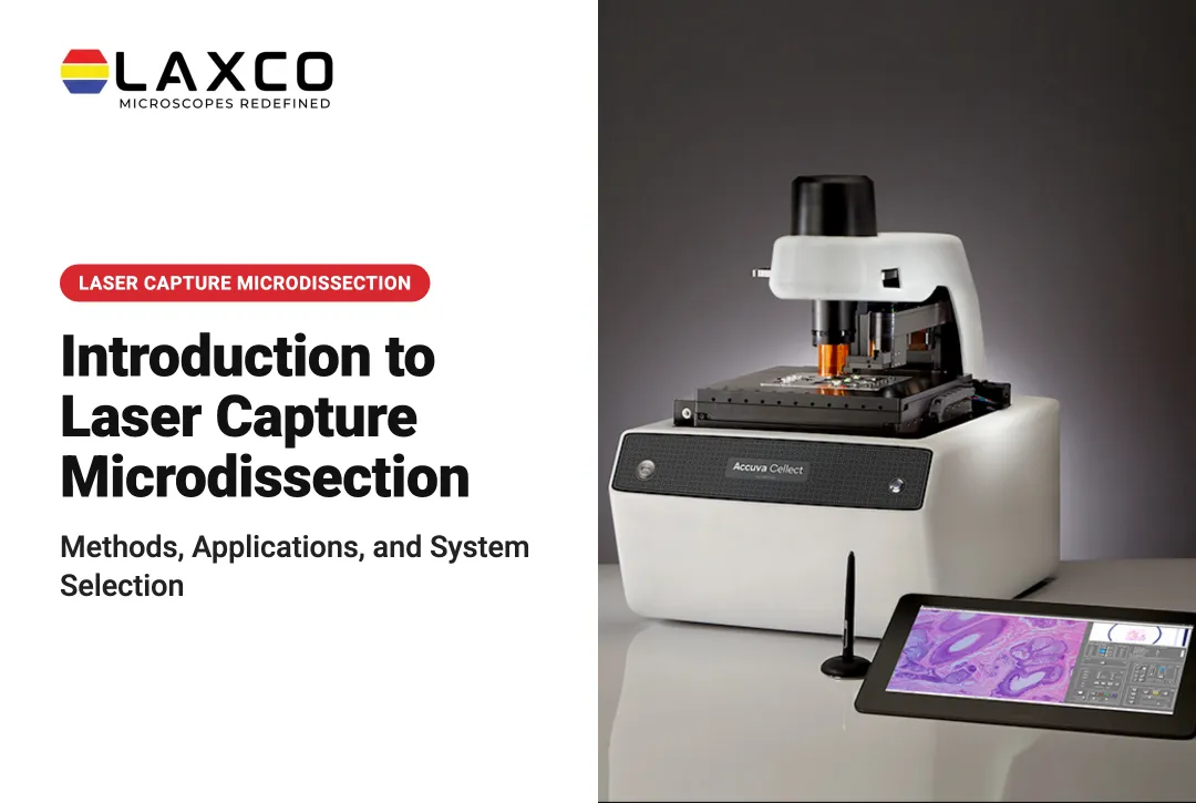
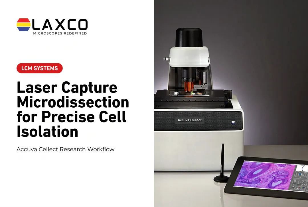
Laxco's Accuva Cellect Laser Capture Microdissection System includes both laser types:
- Infrared (IR) laser: designed for film capture of single cells or small numbers of cells.
- UV laser: designed for cutting dense structures, such as bone or plant tissue.
- Sample preparation: Laxco lists FFPE and fresh-frozen tissue, H&E-stained and LCM-optimized stains, immunofluorescence, unstained samples, and cell culture.
- Software workflow: a five-step workflow with visual quality control.
- Footprint: Laxco describes Accuva Cellect as having a footprint approximately 32% smaller than comparable microscope-and-laser systems. Confirm exact dimensions for the selected configuration before planning bench space.
Laxco also offers LEAP AI for separate microscopy image-analysis requirements. LEAP AI pairs Accuva LEAP optics with Media Cybernetics Image-Pro AI for pre-trained deep-learning segmentation, measurement, and reporting. It is not documented as an integrated Accuva Cellect target-selection or capture-control feature. Any workflow using image annotations from a separate system would require compatible image registration and sample handling.
Laxco lists training and 48-hour response times for Accuva Cellect. Researchers should confirm specific training arrangements and service coverage with the company. Its Cellect Grant Assistance Program provides Grant Language Kits for NIH, NSF, HHMI, and DOD proposals, along with a 15-minute Grant Fit consultation.
Frequently Asked Questions
When is LCM the right isolation method?
LCM suits targets defined by morphology or location in a section, such as tumor regions, specific cell layers, or rare cells. Dissociation-based methods such as FACS suit suspensions where tissue location is not needed.
Is LCM compatible with frozen and paraffin-embedded tissue?
Yes. Fresh-frozen tissue generally gives better RNA quality, while FFPE tissue preserves morphology and allows archival studies. Laxco lists both as supported by Accuva Cellect, along with stained, immunofluorescence, unstained, and cell culture samples.
Which downstream analyses can Accuva Cellect support?
Accuva Cellect isolates selected cells or regions as part of a broader microgenomics workflow. DNA and RNA extraction, protein extraction, amplification, NGS library preparation, qPCR, sequencing, and mass spectrometry are performed separately after capture. The instrument does not perform extraction, qPCR, sequencing, or mass spectrometry itself.
Do I need both IR and UV lasers?
The choice depends on your samples and targets. IR thermoplastic-film capture is designed for single cells and small groups in soft tissue. UV cutting can also isolate single cells when optics and workflow allow, and is used for larger regions and dense tissue such as bone or plant material. Accuva Cellect includes both lasers so the method can be matched to each target.
How much bench space does Accuva Cellect need?
Laxco describes Accuva Cellect as having a footprint approximately 32% smaller than comparable microscope-and-laser systems. Confirm exact dimensions for the selected configuration before planning bench layout, and allow space for the instrument, control equipment, sample preparation, and consumables.