
Key Takeaways
- Spatial biology retains tissue architecture that bulk and dissociated methods remove.
- Nature Methods named spatially resolved transcriptomics its Method of the Year for 2020.
- Spatial transcriptomics and proteomics map molecular features within tissue, while laser capture microdissection (LCM) links downstream molecular analysis to selected, documented tissue regions. Image analysis supports interpretation of these different methods.
- Spatial biomarkers gain meaning from their location and surrounding cells.
- Reproducibility, cost, throughput, and computational demands remain challenges.
What Is Spatial Biology?
Spatial biology connects molecular measurements to the organization and location of cells within tissue. Some methods measure molecules directly in tissue sections, while others, including LCM-based workflows, analyze selected tissue regions after isolation while retaining information about their original location. It includes several related areas:
- Spatial genomics: DNA-level features mapped within tissue.
- Spatial transcriptomics: gene expression mapped by location.
- Spatial proteomics: protein distribution across tissue.
- Spatial multiomics: combined measurement of more than one molecular layer.
Nature Methods named spatially resolved transcriptomics its Method of the Year for 2020 (Nature Methods, 2021), reflecting rapid development of the field.
Why Spatial Context Matters
A protein's function can depend on its neighboring cells, tissue layer, or distance from a blood vessel. Spatial methods reveal:
- Cell-to-cell interactions at tumor margins
- Patterns of immune cell infiltration around lesions
- Organization of neural circuits in brain tissue
A 2025 study in Nature Genetics used high-definition spatial transcriptomics to profile immune cell populations in colorectal cancer (Oliveira et al., 2025). The authors described macrophage subpopulations located in distinct spatial niches.

Spatial data also support research into biomarkers and therapeutic targets that depend on tissue location.
Key Techniques
Spatial Transcriptomics
- In situ hybridization methods use labeled probes to detect and localize RNA molecules in cells. Multiplexed methods use sequential imaging rounds and error-robust barcodes to measure hundreds of RNA species.
- Spatial barcoding methods capture RNA on surfaces with positional barcodes, so sequencing reads can be mapped back to tissue locations.
Spatial Proteomics
- Multiplexed immunofluorescence detects many proteins on one section through repeated staining or barcoded antibodies.
- Mass spectrometry imaging measures molecules directly from tissue and records their positions for comparison with histology.
Laser Capture Microdissection
LCM takes a different approach from in situ imaging methods that produce continuous molecular maps across a tissue section. Instead of measuring molecules in place across the full field, LCM physically isolates selected cells or regions under microscopic guidance so they can be analyzed with downstream sequencing or proteomics workflows.
The resulting measurements are therefore region-based: each isolate is tied to a documented location and morphology, but the molecular data come from the selected material rather than from a continuous spatial assay of the entire section.
LCM is useful when laboratories need deeper or untargeted molecular data from defined regions, when existing sequencing or mass spectrometry resources can be reused, or when the research question centers on selected populations rather than a full-section spatial map. It can complement in situ spatial methods rather than replace them.
Image Analysis
Spatial experiments generate large image datasets, and manual cell segmentation does not scale. Deep-learning methods can segment cells across large sample sets with consistent parameters. Segmentation quality varies with tissue and imaging modality, so methods should be evaluated on the data at hand (Chen and Murphy, 2023).
Spatial Biomarkers and Spatial Gene Expression
Spatial biomarkers are molecular markers whose diagnostic or prognostic value depends on location and surrounding cells, not only on presence.
Spatial gene expression describes which genes are active, and at what level, at specific positions in tissue.
A 2022 study in the Journal for ImmunoTherapy of Cancer used spatially resolved proteomic profiling in advanced non-small cell lung cancer (Moutafi et al., 2022). It identified CD44 on tumor cells as a biomarker associated with sensitivity to PD-1 axis blockade. This illustrates how the cell compartment in which a marker is measured can affect its interpretation.
Challenges and Future Directions
- Reproducibility: sample handling and protocols vary between laboratories.
- Cost: specialized reagents and instruments are expensive.
- Throughput: many workflows are slower than bulk methods.
- Computation: large datasets require substantial processing and expertise.
A 2025 Nature Biotechnology paper proposed standardized metrics for assessing the quality and reproducibility of imaging-based spatial transcriptomics datasets (Plummer et al., 2025).

Directions for the field include:
- Greater automation of sample preparation and imaging
- Higher multiplexing
- Subcellular resolution
- Integration of transcriptomic and proteomic data
Laxco Tools Relevant to Spatial Biology Workflows
Laxco offers instruments that support different stages of tissue selection, microscopy, and image analysis. Their functions should be distinguished rather than presented as one integrated spatial-omics platform.
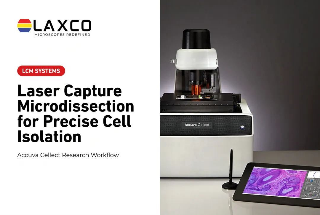
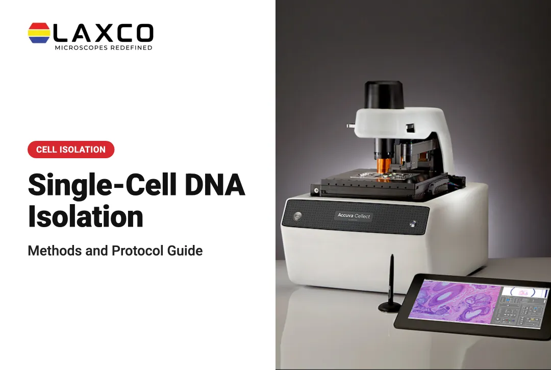
- Accuva Cellect: laser capture microdissection for isolating cells or regions before downstream molecular analysis. An infrared laser supports capture of single cells or small numbers of cells, and a UV laser supports cutting of dense structures. Compatible sample types include FFPE, fresh-frozen, stained, unstained, immunofluorescence, and cell culture. Its footprint is about 32% smaller than comparable microscope-and-laser systems.
- LEAP AI: a separate microscopy and image-analysis platform that pairs Accuva LEAP optics with Image-Pro AI. It is not an integrated Accuva Cellect capture step and is not a spatial transcriptomics assay.
- SLi6Pro: an inverted fluorescence microscope with SeBaLIT illumination across 390-662 nm and 3-color multiplex capability. It does not perform spatial transcriptomics or mass spectrometry spatial proteomics.
These are distinct research tools. They are not automatically connected into a tissue-profiling, sequencing, or spatial multiomics workflow.
Laxco lists training and 48-hour response times for Accuva Cellect. Its instruments support microscopy, tissue isolation, or image analysis according to the product; Laxco does not perform spatial transcriptomics or proteomics as a laboratory testing service.
Frequently Asked Questions
How do I choose between spatial transcriptomics and spatial proteomics?
Choose spatial transcriptomics when the research question concerns RNA expression patterns and the location of gene-expression signals. Some methods provide broad transcriptome coverage, while others measure predefined gene panels. Choose spatial proteomics when protein localization or abundance is the main objective. Protein modifications or activity can be studied only when the chosen assay specifically measures those features. Projects requiring both molecular layers may use compatible multiomic methods or matched serial sections, depending on the platform and sample requirements.
What are spatial biomarkers?
Spatial biomarkers are markers whose significance depends on location and neighboring cells. For example, a 2022 study linked CD44 measured on tumor cells, rather than overall, with response to PD-1 axis blockade in non-small cell lung cancer.
How does laser capture microdissection support spatial biology?
LCM supports spatial biology through region-based sampling rather than a continuous molecular map of the full section. It isolates selected cells or regions under microscopic guidance for downstream sequencing or proteomics while documenting their original location and morphology. It complements in situ methods when laboratories need deeper or untargeted data from defined regions of interest.
Is spatial biology used in clinical diagnostics?
Spatial biology is mainly a research tool. Translational studies are growing, particularly in oncology, but routine clinical use is limited. Laxco's Accuva Cellect and LEAP AI are research instruments, not diagnostic devices.
What should I compare before building spatial capability?
Compare the molecular layer you need, required resolution, tissue types, throughput, and analysis resources. LCM is one option when region-based isolation fits the question and existing sequencing or mass spectrometry resources are available, but it is not a mandatory starting point. Laboratories may begin with imaging-based spatial methods, LCM-enabled workflows, or a combination, depending on their goals and infrastructure.