Key Takeaways
- Spatial proteomics measures protein abundance and location together, at subcellular, cellular, or tissue-region scale.
- Protein mislocalization has been associated with a range of diseases.
- Antibody-based imaging measures predefined protein panels, including methods that use mass spectrometry to detect metal-tagged antibodies. Antibody-free LC-MS/MS can provide broader protein identification from isolated material, while mass spectrometry imaging maps detected molecular signals across tissue.
- Workflows that combine imaging, laser microdissection, and mass spectrometry link protein profiles to defined cell phenotypes.
- Every method involves trade-offs among plex, resolution, throughput, and cost.
What Is Spatial Proteomics?
Spatial proteomics operates at more than one biological scale. At the subcellular level, it maps proteins to organelles and compartments within individual cells. At the tissue level, it maps proteins across cells and regions in a section. A 2019 review in Nature Reviews Molecular Cell Biology described the spatial proteome as protein localization and its dynamics at the subcellular level (Lundberg and Borner, 2019). In tissue research, the same term is used for cell- and region-resolved protein maps.
Methods fall into two broad groups. Antibody-based imaging measures predefined protein panels at high spatial resolution, including fluorescence multiplex panels and mass cytometry methods that detect metal-tagged antibodies. Antibody-free approaches include mass spectrometry imaging, which maps detected molecular signals across a section, and LC-MS/MS after isolation of selected material, which can identify a broader set of proteins from that material. Spatial proteomics therefore asks which proteins are present and where they are, at the scale the chosen method can resolve. Bulk proteomics answers abundance after homogenization and averages across many cells, which can obscure differences between populations in heterogeneous tissue such as tumors with mixed immune and cancer cells.
Why Protein Location Matters
Many proteins act in specific compartments, such as the nucleus, mitochondria, or plasma membrane. Moving a protein between compartments can change its activity. Transcription factors, for example, regulate genes only when they are in the nucleus.
Protein mislocalization has been linked to several inherited and acquired disorders, including defects in nuclear transport, peroxisomal import, and mitochondrial targeting. Resources such as the Human Protein Atlas subcellular section map the subcellular locations of human proteins using antibody-based imaging.
Main Spatial Proteomics Methods
Antibody-Based Multiplexed Imaging
Multiplexed immunofluorescence (IF) uses repeated cycles of antibody staining, imaging, and signal removal or inactivation. Each cycle adds a few markers, and the cycles are combined into a high-plex image. Related fluorescence methods use DNA-barcoded antibodies revealed iteratively. These approaches are targeted: they measure proteins for which validated antibodies exist.
Imaging mass cytometry (IMC) is also antibody-based and targeted, but it labels antibodies with metal isotopes and reads them by mass spectrometry rather than fluorescence. Because fluorophores are not used, spectral overlap is not the limiting factor; acquisition is typically slower than fluorescence imaging, and the tissue is consumed during analysis. A Nature Reviews Methods Primers overview covers multiplexed antibody imaging strategies of this kind (Nature Reviews Methods Primers).
Practical considerations include:
- Each antibody must be validated for the tissue and fixation method.
- Conventional fluorescence typically allows only a few channels per cycle because of spectral overlap.
- High-plex runs require long acquisition and image-registration time.
Mass Spectrometry-Based Approaches
Mass spectrometry can identify proteins without a predefined antibody panel. Two antibody-free spatial strategies are common:
- Mass spectrometry imaging (MSI): measures molecular signals directly across a tissue section and produces a continuous spatial map of detected ions. Protein identification depth is generally lower than in bulk LC-MS/MS workflows.
- Laser capture microdissection (LCM) followed by LC-MS/MS: selected regions or cells are isolated, then analyzed by liquid chromatography-tandem mass spectrometry. This can yield deeper protein identification from defined material, but it is region-based sampling rather than a continuous tissue-wide molecular map (review).
Smaller sampled regions provide finer spatial detail but less material, which usually reduces the number of proteins identified.
Imaging-Guided Microdissection and Mass Spectrometry
Some hybrid workflows combine three steps:
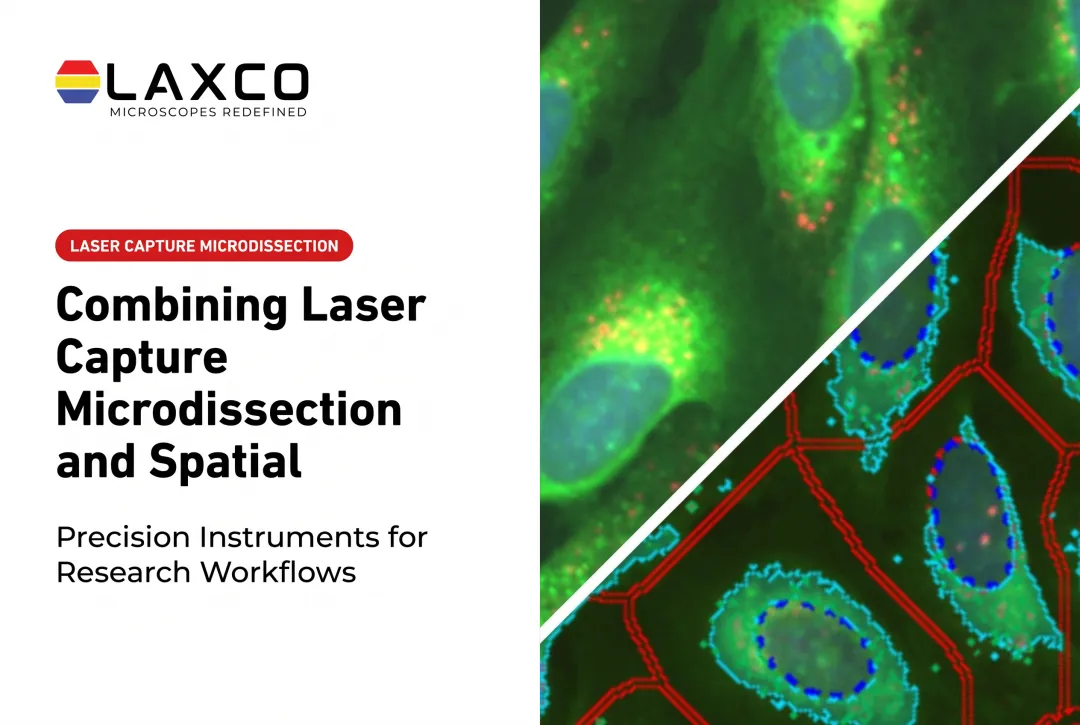
- Image analysis identifies and classifies cells in whole-slide images.
- Laser microdissection isolates the classified cells or regions.
- Mass spectrometry profiles the proteome of the isolated material.
Imaging-guided microdissection connects protein measurements to cells or tissue regions selected through microscopy and image analysis. Researchers can identify targets according to morphology, staining, or validated image-based classifications, isolate the selected material using laser microdissection, and perform protein extraction and mass spectrometry as separate downstream steps.
The accuracy of this approach depends on reliable image analysis, precise region selection, capture quality, and appropriate low-input proteomics methods. The resulting protein data can be associated with the documented location and characteristics of the collected material.
When multiple cells are isolated and analyzed together, the resulting proteomic measurements represent the combined population rather than an independent proteome for every cell. Unlike continuous spatial imaging methods, region-based LCM followed by LC-MS/MS does not automatically produce a pixel-level protein map of the entire tissue section.

Applications
- Cancer research: mapping immune and tumor cell interactions and identifying region-specific protein signatures.
- Neurobiology: profiling protein aggregates, such as amyloid plaques, compared with surrounding tissue.
- Biomarker research: identifying proteins whose location, not only abundance, is associated with disease features.
Findings from small cohorts should be treated as hypothesis-generating until they are validated in larger studies.
Trade-Offs in Spatial Proteomics
No single method maximizes every parameter. Researchers balance:
- Plex: how many proteins are measured at once.
- Resolution: how precisely location is determined.
- Throughput: how much tissue can be processed in a given time.
- Cost: instrument time, reagents, and analysis effort.
Higher plex in antibody imaging requires more validated antibodies and more cycles. Finer spatial sampling for mass spectrometry reduces input and protein coverage.
Laxco Tools for Spatial Proteomics Workflows
Laxco instruments support imaging and isolation steps used in spatial proteomics workflows. They are separate platforms and do not form a single integrated analysis system:
- Accuva Cellect: a laser capture microdissection system with an infrared (IR) laser for capture of single cells or small numbers of cells and a UV laser for cutting dense structures. Laxco lists FFPE and fresh-frozen tissue, several staining types, immunofluorescence, unstained samples, and cell culture as supported. Protein extraction and mass spectrometry are separate downstream laboratory procedures, not functions of the instrument. Laxco lists training and 48-hour response times for Accuva Cellect.
- LEAP AI: combines Accuva LEAP optics with Media Cybernetics Image-Pro AI for pre-trained deep-learning segmentation, measurement, batch processing, and reporting. LEAP AI is a separate imaging and analysis platform; it is not built into Accuva Cellect, does not automatically select cells for Cellect capture, and does not control the Cellect laser.
- SLi6Pro: an inverted widefield fluorescence microscope with SeBaLIT™ LED-based illumination. It supports three-color multiplex fluorescence imaging and lists compatibility with more than 110 fluorochromes across approximately 390–662 nm. Fluorochrome compatibility is not the same as simultaneous channel capacity.
Laxco does not perform proteomic analysis or mass spectrometry as a laboratory service.
Frequently Asked Questions
How is spatial proteomics different from single-cell proteomics?
Single-cell proteomics measures proteins in individual cells, often after dissociation, and typically does not retain tissue coordinates. Spatial proteomics records where proteins are located in intact cells or tissue sections. Some workflows combine both by isolating imaged cells or regions, for example by laser capture microdissection, and analyzing them by LC-MS/MS; that isolation step is region-based and does not automatically create a continuous tissue-wide protein map.
Can I use LCM to prepare samples for mass spectrometry?
Yes. LCM isolates selected cells or regions for protein extraction and LC-MS/MS. Laxco's workflow for Accuva Cellect includes protein extraction and mass spectrometry as downstream options, with the analysis performed in your laboratory.
Which imaging approach suits cultured cells?
Multichannel immunofluorescence on an inverted widefield fluorescence microscope suits many cultured-cell studies. The SLi6Pro supports three-color multiplex fluorescence with SeBaLIT illumination across approximately 390–662 nm and lists compatibility with more than 110 fluorochromes. Treat the dye count as fluorochrome compatibility, not simultaneous channel capacity.
Can AI segmentation guide which cells to isolate?
Segmentation can classify cells by phenotype and help define capture targets for a separate microdissection step. LEAP AI provides pre-trained deep-learning segmentation on a separate imaging platform; it does not automatically transfer selections to Accuva Cellect or control the Cellect laser. Any segmentation used to guide isolation should be validated against annotated images before it is applied to a study.
Which sample types does Accuva Cellect support?
Laxco lists FFPE and fresh-frozen tissue, H&E-stained and LCM-optimized stained sections, immunofluorescence, unstained samples, and cell culture. Staining effects on protein recovery are not uniform across analytes and assays; choose staining and extraction workflows validated for the intended downstream method (Espina et al., 2007).