
Key Takeaways
- Laser microdissection isolates selected cells or regions under direct visualization, reducing collection of neighboring cells.
- Infrared (IR) capture and ultraviolet (UV) cutting suit different targets and tissues.
- Captured material is used for PCR, RNA sequencing, and proteomics.
- Fixation, staining, slide choice, and dehydration affect downstream results.
- System choice depends on laser type, magnification, collection method, and software.
What Is Laser Microdissection?
Laser microdissection combines a microscope with laser optics. The researcher identifies a region of interest on a stained or unstained slide, and the laser captures or cuts it for collection.
General workflow:
- Locate the region of interest on a mounted section.
- Define the target and capture or cut it with the laser.
- Collect the material into a cap, tube, or well plate for extraction.
Laser microdissection can be applied to many sample types, including FFPE sections, frozen tissue, cytology preparations, and cell cultures.
A Note on Terminology
The terms "laser capture microdissection" (LCM) and "laser microdissection" (LMD) overlap in scientific literature and product documentation. Historically, LCM has often been associated with infrared thermoplastic-film capture and LMD with UV laser cutting, but these are not universal definitions. Modern instruments may use IR capture, UV cutting, or both, with different collection mechanisms. When comparing methods, distinguish the actual laser and tissue-collection architecture rather than relying on the terminology alone.
How the Technology Works
Infrared (IR) thermoplastic-film capture: in conventional film-based systems, an IR laser pulse activates the film on a capture surface so it adheres to selected cells or tissue regions. The material is retrieved with that capture surface. The approach commonly supports individual cells or small populations, but collection mechanisms and sample requirements depend on the platform.
Ultraviolet (UV) cutting: a focused UV laser cuts around selected cells or tissue regions, including through a supporting membrane when the workflow uses membrane-mounted sections. The dissected material is recovered by gravity, laser-assisted transfer, or another platform-specific mechanism. UV cutting can isolate individual cells as well as larger regions; suitability for dense tissue depends on the instrument and sample preparation.
Microscope and Collection
LCM systems combine upright or inverted microscopes with laser optics and several objectives: low magnification to locate regions and higher magnification to define targets.
Collection methods include:
- Gravity transfer into a collection vessel
- Laser pressure transfer
- Film-based transfer onto collection caps
Sample Preparation
Fresh-Frozen and FFPE Tissue
Frozen sections generally preserve RNA better than formalin-fixed paraffin-embedded (FFPE) sections. Frozen tissue is typically embedded in OCT, stored at -80°C, and kept cold until sectioning and staining.
FFPE tissue preserves morphology and provides access to archived samples, but fixation and storage can reduce nucleic acid quality. Validate the full workflow with your intended assay before starting a study.
Staining and Slides

Common staining options include:
- H&E for general morphology
- Toluidine blue for rapid contrast on frozen sections
- Immunohistochemistry for marker-specific targeting
- Immunofluorescence for identifying defined cell populations
Membrane slides are used in many UV laser microdissection workflows to support tissue cutting and recovery. However, slide preparation and dehydration requirements depend on the instrument, tissue, preservation method, and collection mechanism. IR thermoplastic-film capture commonly requires appropriately dry tissue for efficient transfer to the capture surface. UV-based systems should follow their validated preparation requirements rather than applying the same dehydration sequence universally.
Research Applications
Cancer research: isolating tumor cells from surrounding tissue supports studies of gene expression, tumor heterogeneity, and therapeutic targets. Glioblastoma studies have combined LCM with RNA sequencing to compare histologically distinct tumor regions (Civita et al., 2019).
Proteomics: LCM-isolated cells and regions can be analyzed by mass spectrometry to produce protein profiles linked to tissue location.
Developmental biology and drug discovery: LCM isolates specific cell types at defined stages to study gene function or drug response.
Other fields:
- Neuroscience: isolating neurons or defined brain regions for RNA analysis
- Forensics: separating cells in mixed biological samples
- Plant biology: profiling gene expression in specific tissue types
- Genomics: preparing enriched starting material for sequencing
Advantages and Limitations
Advantages:
- Precision down to single cells
- Documented morphology and location of each sample
- Compatibility with PCR, sequencing, and mass spectrometry
Limitations:
- Instruments and consumables add cost.
- Operators need training to identify targets and capture cleanly.
- Fixation and storage affect RNA and protein recovery.
- Small inputs may require amplification or highly sensitive downstream methods.
Choosing a System
| Factor | What to consider |
|---|---|
| Laser type | Compare IR film-based capture and UV cutting by supported target size, tissue preparation, cutting capabilities, and collection mechanism. Both can isolate small targets depending on the system. |
| Magnification | Suitable for your smallest targets |
| Collection | Compatible with your extraction tubes or caps |
| Software | Target selection, quality control, and documentation |
| Support | Training and technical support availability |
Laxco Systems
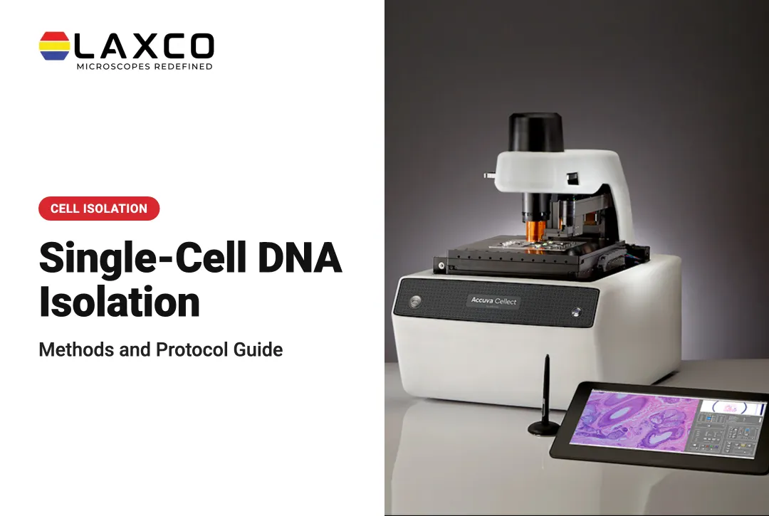
Accuva Cellect, a dedicated laser capture microdissection system:
- Infrared (IR) laser: designed for capture of single cells or small numbers of cells.
- UV laser: designed for cutting dense structures, such as bone or plant tissue.
- Sample preparation: Laxco lists FFPE and fresh-frozen tissue, H&E-stained and LCM-optimized stains, immunofluorescence, unstained samples, and cell culture.
- Collection: compatible with CapSure LCM caps.
- Software workflow: a five-step workflow with visual quality control.
- Footprint: approximately 32% smaller than comparable microscope-and-laser systems, according to Laxco. Confirm the exact dimensions for the selected configuration before planning bench space.
LEAP AI is a separate microscopy and image-analysis platform combining Accuva LEAP optics with Media Cybernetics Image-Pro AI for segmentation, measurement, and batch-ready analysis protocols. It is not a documented built-in feature of Accuva Cellect and should not be described as controlling or automating Cellect's laser-capture workflow.
Laxco lists training and 48-hour response times for Accuva Cellect. Specific training arrangements and service coverage should be confirmed with Laxco. Its Cellect Grant Assistance Program offers Grant Language Kits for NIH, NSF, HHMI, and DOD proposals.
Frequently Asked Questions
Which samples are best suited to laser microdissection?
Laser microdissection suits heterogeneous tissue where target cells are small, scattered, or defined by location. Laxco lists FFPE and fresh-frozen tissue, several stains, immunofluorescence, unstained samples, and cell culture as supported by Accuva Cellect.
Can laser microdissection be used for proteomics?
Yes. Microdissected cells and regions can be analyzed by mass spectrometry, and Laxco's workflow for Accuva Cellect includes protein extraction. Use extraction methods adapted to small inputs and to the fixation used.
Do I need membrane slides?
Membrane slides are commonly used for UV cutting because the membrane supports the excised piece during transfer. IR capture is often performed on glass slides. Choose slides that suit the capture method and tissue type.
How should sections be prepared before capture?
Follow the validated protocol for the tissue, preservation method, LCM platform, and downstream assay. Mount sections on compatible slides and apply staining where needed to identify targets. IR thermoplastic-film capture commonly requires appropriately dehydrated, dry sections for reliable film transfer, while UV cutting workflows may have different preparation requirements. Do not apply one universal ethanol-dehydration sequence to every LCM method.
What support does Laxco provide for new LCM users?
Laxco lists training and 48-hour response times for Accuva Cellect. Researchers should confirm specific training formats, service terms, and application assistance with a Laxco specialist. The Cellect Grant Assistance Program also provides technical information and Grant Language Kits for funding proposals.