Key Takeaways
- Laser microdissection isolates selected cells or regions while documenting their morphology and location.
- Infrared (IR) film-based capture and ultraviolet (UV) laser cutting use different isolation and collection mechanisms. Both can isolate small targets, including individual cells, while suitability for larger or dense regions depends on the instrument and tissue preparation.
- A typical workflow includes sample preparation, target identification, dissection and collection, and molecular analysis.
- Applications include oncology, neuroscience, plant biology, and imaging-guided proteomics.
- System choice depends on sample type, target size, throughput, and downstream assay.
What Is Laser-Assisted Microdissection?
Laser microdissection combines a microscope with a laser to separate targets from a tissue section. The researcher views the section, marks the cells or region of interest, and activates the laser to isolate that material.
Tissue sections often contain several cell types, such as tumor cells, immune cells, connective tissue, and blood vessels. Extracting nucleic acids or proteins from the whole section produces an average signal. Microdissection enriches the population of interest before extraction.
Emmert-Buck and colleagues at the National Institutes of Health described laser capture microdissection in 1996. They presented it as a method for procuring pure cell populations from tissue for molecular analysis (Emmert-Buck et al., 1996).
Main Techniques
Infrared Laser Capture
In conventional IR thermoplastic-film capture, a film-coated capture surface, often incorporated into a collection cap, is positioned against the tissue section. A focused infrared laser pulse locally activates the thermoplastic film so it adheres to selected cells or tissue regions. The target material is then lifted from the slide with the capture surface. This method is commonly used for individual cells or small cell populations. Collection mechanics and suitable sample preparations depend on the instrument.
UV Laser Cutting
In UV laser microdissection, a focused ultraviolet laser cuts around a selected cell or tissue region. Depending on the system, the isolated material may be collected by gravity, laser-assisted transfer, or another compatible collection mechanism. UV cutting can isolate individual cells as well as larger regions, and certain systems are suitable for dense tissue structures. The supported target sizes and tissue types depend on the instrument and workflow.
| Feature | IR film-based capture | UV laser cutting |
|---|---|---|
| Isolation principle | Laser-activated thermoplastic film adheres to selected material | Focused UV laser cuts around selected cells or tissue regions |
| Target size | Individual cells or small cell populations; depends on capture configuration | Individual cells, small populations, or larger regions; depends on system configuration |
| Collection | Selected material is lifted with the film-coated capture surface | Cut material is recovered using the platform's collection mechanism |
| Selection considerations | Film compatibility, tissue preparation, and capture efficiency | Cutting precision, tissue properties, collection method, and downstream assay |
Some systems combine both lasers so that researchers can match the method to each target.

Typical Workflow
- Prepare the sample. Section and mount the tissue using the fixation, preservation, and slide conditions required by the instrument and downstream assay. Where staining is needed, use an appropriate H&E, immunofluorescence, or LCM-optimized protocol. Some workflows also support unstained sections.
- Identify the target. Mark the cells, region, or structure of interest in the software.
- Dissect and collect. Use the laser to capture or cut the target and transfer it to a cap, tube, or plate.
- Extract and analyze. Lyse the material and extract DNA, RNA, or protein for amplification, sequencing, or mass spectrometry.
The amount of material required depends on preservation method, target molecule, and assay sensitivity. There is no single universal cell-count requirement, so pilot experiments are useful.
Research Applications
Cancer Research
Separating tumor-rich regions from surrounding stroma or other tissue compartments can reduce dilution of mutations and gene-expression signals by unrelated cell populations. Researchers can use laser microdissection to isolate histologically defined areas, such as tumor, adjacent tissue, and vascular regions, before performing separate DNA or RNA analyses.
The resulting molecular profiles represent the collected cells or regions. When multiple cells are captured together, these measurements describe the combined population rather than independent single-cell profiles.
Neuroscience
Specific neuronal populations can be isolated from brain sections for expression profiling. This is useful where neighboring cell types differ markedly in gene expression.
Plant Biology
Plant cell walls make manual separation of individual cell types difficult. Laser microdissection allows specific plant tissues and cell types to be collected for RNA sequencing.
Imaging-Guided Proteomics
Imaging-guided proteomics combines microscopic tissue examination, targeted laser microdissection, and downstream mass spectrometry to investigate proteins in selected cells or tissue regions.
Researchers can identify regions using tissue morphology, staining, or compatible image-analysis methods, isolate the selected material, and analyze its proteins using separately performed extraction, digestion, and mass-spectrometry workflows.
This approach connects proteomic measurements to the documented location of the collected material. However, analyzing microdissected regions does not automatically produce a continuous spatial protein map or establish individual single-cell protein profiles when multiple cells are collected together.
Choosing a Laser Microdissection System
Sample compatibility: confirm support for your sample types, such as frozen sections, FFPE tissue, cell cultures, and dense materials like bone or plant tissue.
Target size: both IR film-based capture and UV cutting can isolate small targets, including individual cells, depending on the system. Evaluate the collection mechanism, supported tissue types, capture efficiency, and ability to isolate the required target size. For dense structures, confirm the instrument's specific cutting capabilities rather than relying on laser type alone.
Throughput: laboratories processing many samples benefit from software that streamlines target selection and documentation.
Downstream compatibility: collected material should be compatible with the planned assay, whether PCR, sequencing, or mass spectrometry.
Laxco Accuva Cellect
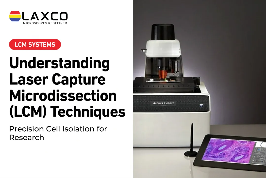
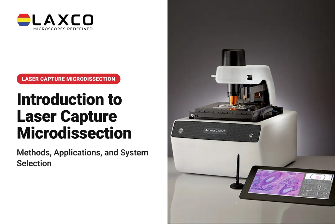
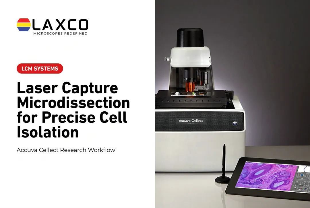
Laxco's Accuva Cellect Laser Capture Microdissection System combines both laser types in one instrument:
- Infrared (IR) laser: designed for capture of single cells or small numbers of cells.
- UV laser: designed for cutting dense structures, such as bone or plant tissue.
- Sample preparation: Laxco lists FFPE and fresh-frozen tissue, H&E-stained and LCM-optimized stains, immunofluorescence, unstained samples, and cell culture.
- Software workflow: a five-step workflow with visual quality control before capture.
- Downstream workflows: captured material can proceed to DNA or RNA extraction for analyses such as qPCR or sequencing, or to protein extraction for mass spectrometry. These analytical procedures are separate from the microdissection instrument.
Laxco lists training and 48-hour response times for its Accuva Cellect Laser Capture Microdissection System. The Cellect Grant Assistance Program offers Grant Language Kits for NIH, NSF, HHMI, and DOD proposals.
Frequently Asked Questions
When should I use IR capture rather than UV cutting?
Both IR film-based capture and UV laser cutting can isolate individual cells or small tissue regions, depending on the instrument. IR film-based capture uses a laser-activated surface to retrieve selected material, while UV cutting separates targets along a defined boundary before collection. Laxco's Accuva Cellect positions its IR laser for single cells or small populations and its UV laser for dense structures such as bone and plant tissue. Choose the method according to the sample, collection requirements, and downstream assay.
How much precision does laser microdissection add over manual dissection?
Laser microdissection selects targets under magnification and defines boundaries with the laser, so small or scattered cells can be collected with less neighboring tissue. Manual dissection remains practical for large, well-defined regions.
Which sample preparations does Accuva Cellect support?
Laxco lists FFPE and fresh-frozen tissue, H&E-stained and LCM-optimized stained sections, immunofluorescence, unstained samples, and cell culture. Confirm that the preparation is compatible with your downstream DNA, RNA, or protein extraction.
Can laser microdissection affect the quality of captured biomolecules?
Yes. Molecular quality depends on tissue preservation, fixation, staining, laser settings, capture conditions, handling time, and downstream processing. Optimize instrument settings on representative test sections using the manufacturer's operating guidance. Where UV cutting is used, define cutting boundaries that minimize unwanted damage to the target while retaining the required material. Follow the validated collection, stabilization, extraction, or storage procedure for the analyte being studied rather than applying one universal handling rule to all laser modes.
Does Laxco help researchers fund an LCM system?
Yes. Laxco's Cellect Grant Assistance Program offers Grant Language Kits with technical specifications and a 15-minute Grant Fit consultation for researchers including Accuva Cellect in NIH, NSF, HHMI, or DOD proposals.