Key Takeaways
- Plan equipment around five categories: amplification and analysis, imaging, separation, sample preparation and storage, and sterilization.
- Microscopy supports daily work, from routine checks to quantitative imaging and cell isolation.
- Selection should reflect research scale, budget, compatibility, and vendor support.
- Validate instrument performance at your lab's actual working conditions.
Core Molecular Biology Equipment

Amplification and Analysis Instruments
Thermal cyclers and real-time PCR (qPCR) instruments support nucleic-acid amplification and detection. Conventional PCR amplifies DNA, while qPCR monitors DNA amplification in real time using fluorescent detection.
When RNA is the starting material, a reverse-transcription step converts RNA into complementary DNA (cDNA) before PCR amplification. Reverse-transcription quantitative PCR (RT-qPCR) is commonly used to measure RNA-derived targets, including gene-expression studies, with appropriate normalization and controls.
Other core instruments include:
- Gel electrophoresis systems, which separate DNA, RNA, and proteins by size or charge. Match the power supply to the tank and assay.
- Capillary electrophoresis analyzers, which range from benchtop units to high-throughput systems for fragment analysis and Sanger sequencing.
- Spectrophotometers and microplate readers, which quantify nucleic acids and proteins. Microvolume models measure small sample volumes directly.
Centrifugation and Liquid Handling
Centrifuges separate cellular components and nucleic acids. Relative centrifugal force (g), rotor capacity, tube format, and refrigeration matter more than maximum rpm alone.
Automated liquid handlers can improve precision over manual pipetting, particularly for repetitive transfers. Performance varies with volume and liquid type, so validate accuracy at your working volumes before relying on automation.
Microscopy and Imaging
Microscopes allow researchers to visualize cell structures, protein localization, and tissue organization. Instrument choice has a large effect on image quality and downstream analysis.
Comparing Microscope Types
| Type | Commonly used for | Main trade-off |
|---|---|---|
| Widefield fluorescence | Labeled structures and colocalization | Resolution limited by diffraction to roughly 200–250 nm |
| Confocal | Optical sectioning of thicker samples | Slower acquisition; more light exposure for live cells |
| Transmission electron microscopy | Nanometer-scale subcellular detail | Extensive preparation; no live imaging |
Laxco's inverted fluorescence microscopes use patented SeBaLIT™ illumination:
- The SLi3Pro supports DAPI, FITC, and TRITC fluorochromes.
- The SLi6Pro covers 390–662 nm and supports more than 110 fluorochromes, including CFP, YFP, and RFP.
These configurations allow common labels for nuclei, mitochondria, and actin to be imaged in the same sample.

Laser Capture Microdissection
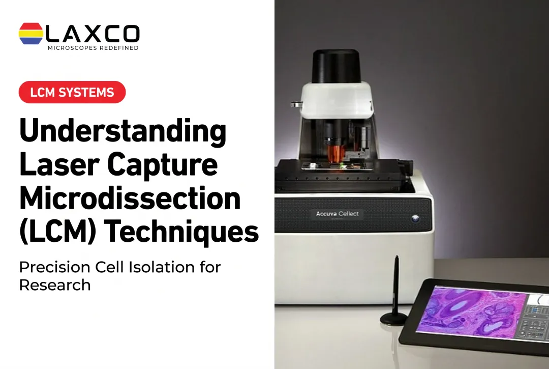
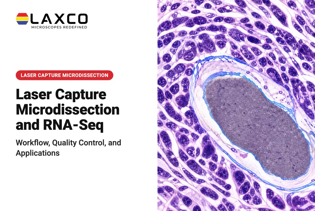
Laser capture microdissection (LCM) isolates selected cells or regions from tissue sections for genomic, transcriptomic, or proteomic analysis. It is useful when surrounding tissue would dilute the signal of interest.
Laxco's Accuva Cellect Laser Capture Microdissection System provides:
- An infrared (IR) laser designed for film capture of single cells or small numbers of cells.
- An ultraviolet (UV) laser designed for cutting dense structures, such as bone or plant tissue.
- A five-step software workflow with visual quality control and documentation.
- A compact footprint that Laxco describes as approximately 32% smaller than comparable microscope-and-laser systems, helping laboratories conserve bench space.
- Documented support for FFPE and fresh-frozen tissue, with downstream use in qPCR, NGS library preparation, and mass spectrometry.
AI-Assisted Image Analysis
AI is increasingly used for image denoising, segmentation, and high-throughput analysis. Validation and reproducibility remain important considerations for any AI-based method.
Laxco's LEAP AI pairs Accuva LEAP optics with Media Cybernetics Image-Pro AI. It provides:
- Pre-trained deep-learning models for AI-assisted segmentation and measurement.
- An Image → Process → Measure → Report workflow.
- Batch-processable life-science protocols with no-code options.
Applying the same validated protocol across operators can reduce variability between users.
Digital All-in-One Microscopes
Digital microscopes integrate camera, computer, and display, which simplifies teaching and shared review. Laxco's SeBa digital microscopes include:
- SeBa2, with touchscreen viewing, HDMI output, and image saving to USB or the SeBaShare cloud.
- SeBaPro4 Series, which integrates conventional microscopy with digital viewing and multi-view options.
Live-Cell Imaging
For time-lapse imaging inside a CO₂ incubator, Laxco's CellLive systems support scheduled acquisition without moving cultures out of the incubator.
Specialized Equipment for Molecular Analysis
Sequencing. Sanger sequencing reads one fragment per reaction and suits confirmation of small targets. Next-generation sequencing (NGS) reads millions of fragments in parallel and suits gene panels, exomes, and whole genomes. Total project cost depends on library preparation and bioinformatics as well as instrument time.
High-performance liquid chromatography (HPLC) and mass spectrometry (MS). HPLC separates compounds based on their interactions with the mobile and stationary phases. Mass spectrometry measures ions according to their mass-to-charge ratio.
The two technologies can be coupled in liquid chromatography-mass spectrometry (LC-MS or LC-MS/MS) workflows, where chromatographic separation precedes mass-based detection. These combined methods are widely used in proteomics, metabolomics, and other analytical applications.
High-throughput screening (HTS). Automated screening systems test large compound libraries in multi-well plates. Real throughput depends on the full workflow, including liquid handling, incubation, and readout.
Cell Culture, Storage, and Sterilization
Cell Culture Equipment
CO₂ incubators control temperature, humidity, and CO₂ concentration for cell growth, commonly at 37 °C. Biological safety cabinets protect against contamination. Class II and III cabinets protect the sample as well as personnel and the environment.
Sample Storage and Preparation
- Ultra-low-temperature freezers preserve nucleic acids, proteins, and cell stocks.
- Cryogenic storage supports long-term preservation of cell lines.
- Vortex mixers, heat blocks, tubes, flasks, and plates support daily preparation.
- Preventive maintenance schedules protect stored samples from equipment failure.
Sterilization
Autoclaves are the standard for sterilizing tools, media, and consumables. Cycle time and temperature depend on the autoclave type, load size, and wrapping. Validate cycles with biological or chemical indicators according to your institution's procedures.
How to Choose Molecular Biology Equipment
- Assess research scale. High-throughput labs need automation and batch capacity. Smaller labs often benefit from versatile instruments.
- Balance quality and budget. Consider warranty coverage, quality-control processes, and technical support alongside purchase price.
- Check compatibility and upgrade paths. This is especially important for software-linked imaging and analysis systems.
- Plan around funding. Laxco's Cellect Grant Assistance Program provides Grant Language Kits for NIH, NSF, HHMI, and DOD proposals that include LCM.
Laxco's in-house quality-control team inspects each microscope during manufacturing and assembly. Checks cover optical clarity, mechanical alignment, and electronic function. For Accuva Cellect, support includes on-site and virtual applications training, live technical support, and a 48-hour response time.
Frequently Asked Questions
Which Laxco microscope suits multicolor fluorescence?
The SLi3Pro supports DAPI, FITC, and TRITC. The SLi6Pro covers 390–662 nm and supports more than 110 fluorochromes. Share your fluorophore list with a Laxco specialist to confirm filter compatibility.
Why is laser capture microdissection used in molecular biology?
LCM isolates selected cell populations from tissue sections, which reduces dilution from surrounding tissue. The isolated material supports DNA, RNA, and protein analysis in cancer and developmental biology research.
What should a new lab prioritize when buying equipment?
Versatile instruments that cover several applications are often a good starting point. Confirm warranty terms, service coverage, training, and quality-control processes before purchasing.
How often should microscopes and centrifuges be maintained?
Follow each manufacturer's maintenance schedule. Routine cleaning, calibration checks, and periodic professional service help maintain accuracy and extend instrument life.