Key Takeaways
- Single-cell methods can detect cell populations and states that bulk measurements obscure.
- Isolation methods differ in throughput, input requirements, and whether tissue context is retained.
- Genomic, transcriptomic, epigenomic, and proteomic measurements are now possible at single-cell resolution, with different depth and coverage.
- Data analysis and integration require specialized computational expertise.
What Is Single-Cell Analysis?
Single-cell analysis measures molecular features of individual cells rather than averaging across a population. A bulk measurement from a tumor biopsy, for example, combines signals from tumor cells, stromal cells, and immune cells. A small subpopulation may contribute too little signal to detect.
Examples of single cells studied in research include:
- A circulating tumor cell isolated from a blood sample.
- A neuron from a defined cortical region.
- A T cell from a tumor microenvironment.
Single-Cell Isolation Techniques
Several approaches are used to obtain individual cells or defined small populations:
- Fluorescence-activated cell sorting (FACS): a flow-based sorting method that selects cells from a suspension using fluorescence and light-scatter signals.
- Microfluidics: handles cells in nanoliter-scale channels or droplets, supporting high-throughput capture with small volumes.
- Manual micromanipulation: picks individual cells under a microscope; low throughput, but useful for cultured cells or embryos.
- Hydrodynamic traps: capture cells in arrays for time-resolved observation.
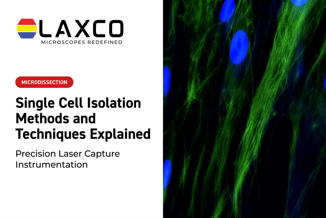
- Laser capture microdissection (LCM): isolates selected cells or regions directly from tissue sections under microscopic guidance, retaining information about tissue location.
Laser Capture Microdissection
LCM does not require dissociating the tissue. The operator identifies target cells in a stained or unstained section. An infrared laser can bond the selected cells to a thermoplastic film for film capture, and a UV laser can cut around a region of interest. The captured material is then processed for DNA, RNA, or protein analysis.
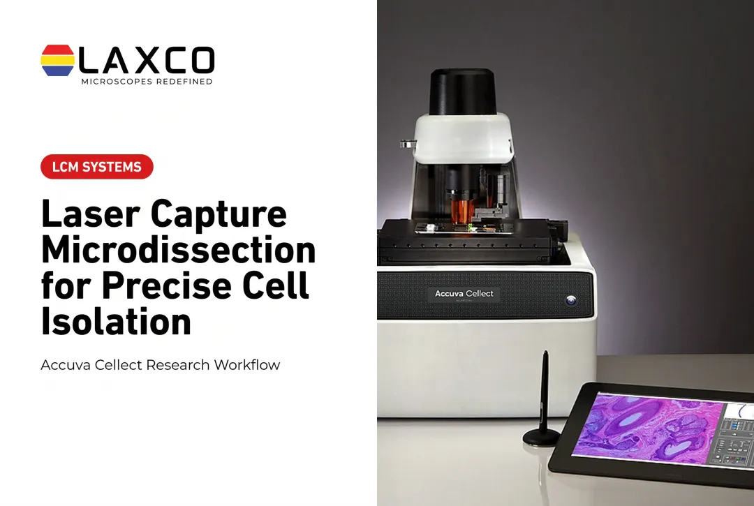
Laxco's Accuva Cellect Laser Capture Microdissection System combines both laser functions:
- An infrared (IR) laser designed for film capture of single cells or small numbers of cells.
- An ultraviolet (UV) laser designed for cutting dense structures, such as bone or plant tissue.
- A five-step software workflow with visual quality control and documentation.
- Documented support for FFPE and fresh-frozen samples, H&E-stained and LCM-optimized stains, immunofluorescence, unstained samples, and cell culture.
Support for FFPE material allows researchers to work with archived tissue collections.
Choosing an Isolation Method
| Method | Commonly used for | Limitation |
|---|---|---|
| FACS | Marker-based sorting of many cells | Requires dissociation; tissue context is lost |
| Microfluidics | High-throughput capture for sequencing | Requires dissociation; some capture events contain more than one cell |
| Hydrodynamic traps | Time-resolved observation of single cells | Limited scale |
| LCM | Morphology- or location-guided selection from tissue sections | Lower throughput; operator-guided |
| Micromanipulation | Live cells and embryos | Slow and skill-dependent |
The choice depends on sample type, cell size, required cell numbers, and the downstream assay.

Core Single-Cell Omics Technologies
Genomics and Whole-Genome Amplification
Single-cell genome sequencing requires library-preparation methods that work with very small amounts of DNA. Whole-genome amplification (WGA) is commonly used to generate sufficient material for sequencing, but it is not required by every method. For example, Zahn et al. (2017) demonstrated single-cell whole-genome library preparation without a preamplification step for copy-number analysis. Where WGA is used, common methods include:
- DOP-PCR: lower genome coverage, used for large-scale copy-number analysis.
- Multiple displacement amplification (MDA): higher coverage through isothermal amplification, with amplification bias that can affect copy-number estimates.
- MALBAC: quasi-linear preamplification designed to reduce amplification bias. In the original study, Zong et al. (2012) reported up to approximately 93% genome coverage at a minimum of one read per position, using a mean sequencing depth of approximately 25× for a single human cell. This was a study-specific result rather than a guaranteed coverage level for every sample.
In a 2011 study, Navin and colleagues used single-nucleus sequencing to investigate copy-number differences within breast tumors. Their analysis of 100 individual nuclei from one tumor revealed distinct genomic subpopulations consistent with sequential clonal expansions, demonstrating how single-cell measurements can resolve tumor heterogeneity that is difficult to distinguish in bulk samples.
Transcriptomics
Single-cell RNA sequencing (scRNA-seq) methods trade transcript coverage against cell throughput:
- Plate-based full-length methods (such as Smart-seq2): capture more of each transcript, supporting splice-variant analysis, at lower throughput.
- Droplet-based methods (such as Drop-seq): encapsulate cells with barcoded beads, enabling thousands of cells per experiment.
Epigenomics and Proteomics
Single-cell ATAC-seq maps chromatin accessibility in individual cells. Other single-cell methods profile histone modifications. These approaches describe regulatory differences between cells that bulk chromatin assays average together.
Single-cell proteomics by mass spectrometry is also feasible. The SCoPE2 method uses isobaric barcoding. A study profiling 1,490 monocytes and macrophages reported about 1,000 proteins quantified per cell and more than 3,000 proteins across the dataset (Genome Biology, 2021).

Spatial Approaches
Spatial methods measure molecular features while retaining their position in tissue:
- Imaging-based methods: multiplexed fluorescence in situ hybridization techniques, including MERFISH, measure predefined RNA targets while preserving their positions within cells and tissue. Panel sizes depend on the assay design and can range from hundreds to thousands of genes. A 2019 MERFISH study (PMC6765259) demonstrated spatial measurements of approximately 10,000 genes in individual cells.
- Sequencing-based methods: capture RNA on spatially barcoded surfaces and sequence it, with resolution depending on the platform.
- Tissue-isolation methods: LCM removes selected regions for downstream molecular analysis, including mass spectrometry.
These approaches differ in how spatial information is generated. Imaging and spatial transcriptomics methods measure molecular signals at known positions within tissue, with resolution determined by the platform. LCM instead selects cells or tissue regions according to their location and morphology, then collects the material for separate molecular analysis. The resulting measurements retain a record of the sampled region but do not inherently produce a continuous tissue-wide molecular map. Researchers should compare these methods by spatial resolution, molecular coverage, input requirements, and throughput.
Research Applications
Cancer Research
Single-cell sequencing is used to describe clonal structure and heterogeneity within tumors. Studies have reported multiple coexisting subclones within individual tumors, which bulk sequencing does not resolve directly.
Drug Response and Immunotherapy Research
scRNA-seq has been used to study cell states associated with therapy response. In metastatic melanoma, a 2018 study by Sade-Feldman and colleagues associated TCF7-positive CD8 T cells with response to checkpoint immunotherapy. These findings are associations within the studied cohorts.
Machine-learning methods are also applied to single-cell data. scDEAL uses transfer learning to predict drug response from scRNA-seq data. Across six datasets and five drugs, it reported a mean AUROC of 0.898 (Nature Communications, 2022). This is retrospective benchmarking, not clinical validation.
Developmental Biology and Neuroscience
Pseudotime methods, such as Monocle, order single cells along inferred developmental trajectories. In neuroscience, Tasic and colleagues profiled roughly 24,000 cells from two mouse cortical regions to define transcriptomic cell types.
Image Analysis for Single-Cell Imaging
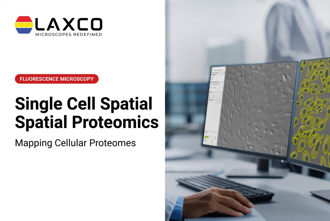
Manual cell segmentation is time-consuming and can vary between analysts. Automated segmentation can reduce manual workload and some sources of analyst-to-analyst variability when validated for the dataset.
Laxco's LEAP AI combines Accuva LEAP optics with Media Cybernetics Image-Pro AI. It supports image acquisition, AI-assisted segmentation, measurement, and reporting through an Image → Process → Measure → Report workflow. It includes pre-trained deep-learning models, batch processing, and no-code protocol options.
Challenges and Considerations
- Cost and expertise: single-cell studies require specialized reagents, instrumentation, and bioinformatics skills.
- Data integration: combining single-cell and bulk data, or multiple omics layers, remains methodologically difficult.
- Technical noise: dropout, batch effects, and amplification bias require careful design and quality control.
- Funding documentation: grant applications usually need a clear rationale for single-cell and LCM methods. Laxco's Cellect Grant Assistance Program provides technical documentation for LCM-based proposals.
Frequently Asked Questions
When should I choose LCM instead of FACS for single-cell work?
LCM may suit studies where cells must be selected by morphology or tissue location, including FFPE samples. FACS may suit high-throughput, marker-based sorting of dissociated cells. The downstream assay and required cell numbers also influence the choice.
Can Accuva Cellect capture a single cell?
Accuva Cellect's IR laser is designed for film capture of single cells or small numbers of cells. Whether single-cell input is sufficient depends on the downstream assay and its sensitivity.
Which sample types does Accuva Cellect support?
Laxco lists FFPE and fresh-frozen tissue, H&E-stained and LCM-optimized stained sections, immunofluorescence, unstained samples, and cell culture. Confirm compatibility with your extraction method during workflow planning.
Does LEAP AI require programming experience?
LEAP AI includes no-code workflow options and pre-trained deep-learning models. Segmentation results should be validated for each sample type and staining approach.