Key Takeaways
- LCM isolates defined cells or regions before protein extraction, so results can be linked to tissue location and morphology.
- LCM combined with LC-MS/MS generates protein profiles from selected, anatomically defined tissue regions rather than continuous pixel-level protein maps. Depending on sample input and analytical methods, it can support deep proteome coverage of those regions.
- Staining helps identify target regions, but its effects on protein and peptide recovery depend on the stain, tissue preparation, and extraction protocol.
- Low input amounts make samples vulnerable to surface losses and contamination.
- Automated sample preparation can reduce variability and processing time for microdissected samples.
Why Combine LCM With Proteomics?
LCM uses a laser to isolate selected cells, structures, or regions from a tissue section under direct visualization. The researcher marks the region of interest, and the laser captures or cuts it.
Homogenizing whole tissue mixes cell types and compartments, which can mask region-specific differences. Enriching the region of interest before extraction preserves information about where proteins are located in tissue architecture.
Comparison With Imaging Mass Spectrometry
Imaging mass spectrometry measures molecular signals across tissue locations to generate spatially resolved images. The measurable molecular classes, spatial resolution, and identification depth depend on the particular method, instrument, sample preparation, and analytical workflow.
LCM combined with LC-MS/MS follows a different approach: defined cells or tissue regions are physically isolated before protein extraction, digestion, and mass-spectrometric analysis. The resulting data describe the proteins recovered from those collected regions rather than producing a continuous protein-abundance map across the original section.
With suitable sample input and highly sensitive analysis, LCM-LC-MS/MS can provide extensive protein identification and quantification for selected regions. However, the achievable depth is assay-dependent and cannot be assumed to exceed every imaging mass-spectrometry method in all experimental conditions.
The Combined Workflow
- Sectioning and mounting: tissue is cut into thin sections and mounted on slides suitable for microdissection.
- Staining and region identification: the researcher or pathologist identifies target regions under the microscope.
- Microdissection and collection: the laser isolates the target cells or region for collection.
- Lysis and digestion: captured material is solubilized and digested into peptides.
- Chromatography and mass spectrometry: peptides are separated and analyzed.

Staining Trade-Offs
Staining helps researchers identify the cells or tissue regions to isolate, but its compatibility with downstream proteomics depends on tissue preservation, staining chemistry, processing conditions, and the protein-extraction method. For FFPE samples, fixation-related modifications and extraction inefficiency can complicate peptide recovery, while suitable retrieval and sample-preparation methods can improve results.
A 2025 review of mass-spectrometry-based FFPE proteomics discusses the influence of tissue preparation, extraction chemistry, and downstream analytical workflows on proteomic data quality.
Researchers should select staining and extraction procedures that preserve the required morphological detail while meeting the validated requirements of their proteomics method. Findings from a particular tissue preparation or laboratory workflow should not be treated as performance specifications for Accuva Cellect.
Automated Sample Preparation
Low-input proteomics is especially sensitive to sample losses during transfer, extraction, digestion, and cleanup. Reducing unnecessary handling and using appropriately validated small-volume or automated sample-preparation methods can help improve consistency.
Automation can be useful after microdissection, particularly when laboratories process numerous region-specific samples. However, the benefits depend on the chosen digestion, cleanup, chromatography, and mass-spectrometry workflow. A 2025 laser capture microdissection methods primer discusses the wider role of sample preparation and downstream analysis in spatially resolved molecular research.
For Laxco's Accuva Cellect, tissue selection and collection are the instrument's documented functions. Automated protein preparation, digestion, chromatography, and mass spectrometry require separately selected downstream equipment and validated laboratory procedures. They should not be presented as integrated Cellect capabilities.
Controlling Contamination
At very low input, contaminating proteins can make up a large share of identifications. Process blanks help distinguish sample proteins from background. In cell culture models, metabolic isotope labeling can also separate labeled sample proteins from unlabeled contaminants.
Applications
Cancer Research
LCM proteomics can compare tumor cells with adjacent stroma or immune cells, and study proteins at specific invasive structures. This links protein changes to defined tissue locations.
Developmental Biology
Microdissection allows individual structures in developing organs to be profiled across time points, rather than averaging the whole organ.
Drug Discovery
Region-specific protein data can identify candidate targets expressed in diseased areas and assess whether they are absent from surrounding normal tissue.
Archival FFPE Tissue
Formalin-fixed paraffin-embedded (FFPE) tissue is widely represented in archival pathology collections. Adapted protein-extraction methods, often involving heat-assisted retrieval and suitable detergents or denaturants, allow proteomic analysis of microdissected FFPE regions. Recovery depends on tissue preservation and extraction conditions, and fixation-related chemical changes may not be completely reversed.
Key Challenges
- Mass-limited samples: small inputs are vulnerable to adsorption on tube surfaces. Reduce volumes and transfer steps.
- Contamination: include process blanks and monitor common contaminants.
- Region selection: the selected boundary determines what the data can show. Document selection criteria.
- Throughput: manual identification and dissection take time. Image analysis and automated preparation can reduce hands-on effort.
Choosing an LCM System for Proteomics
Look for:
- Optics that allow accurate identification of target regions
- Laser options for single cells and for dense tissue
- Reproducible collection with minimal transfer steps
- Compatibility with downstream protein extraction and mass spectrometry
- Software that documents selected regions and quality control
Accuva Cellect for LCM Proteomics
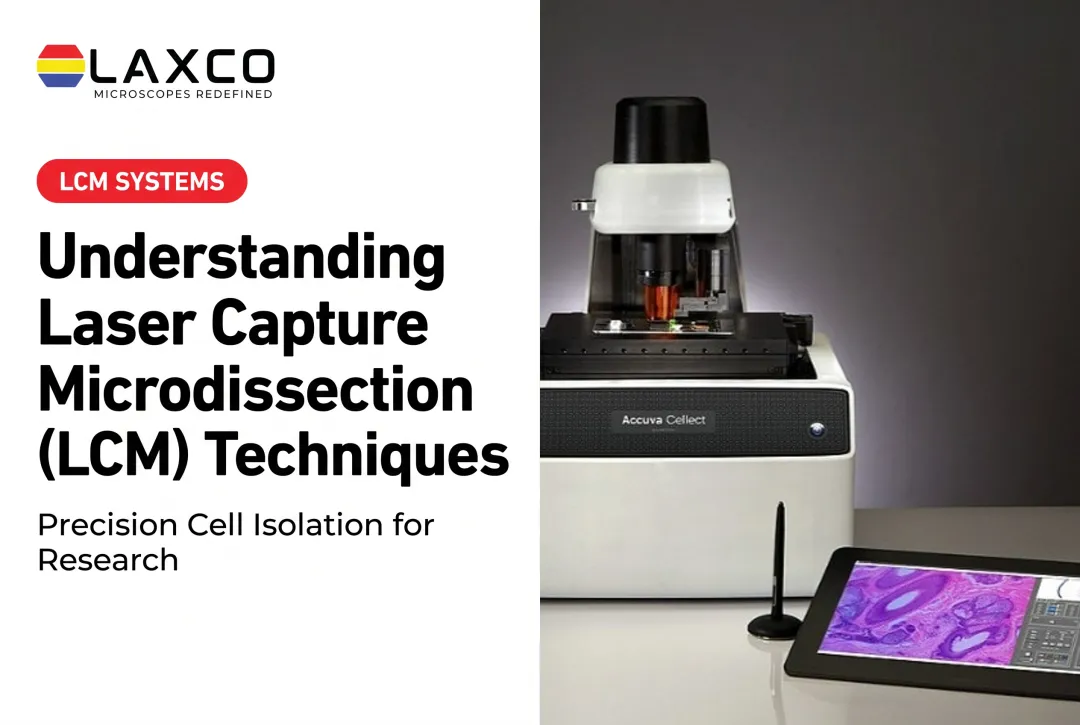
Laxco's Accuva Cellect Laser Capture Microdissection System enables researchers to isolate selected cells or tissue regions before downstream protein extraction and mass-spectrometry analysis. Its IR laser is designed for capturing single cells or small cell populations, while its UV laser supports cutting dense structures. Laxco lists FFPE and fresh-frozen tissue, stained and unstained sections, immunofluorescence, and cell culture among its supported preparations.
The instrument performs tissue selection and collection; protein extraction, digestion, and mass-spectrometry analysis are separate downstream steps. Laxco's LEAP AI is a different microscopy and image-analysis platform, not a documented integrated Accuva Cellect capture feature.
Accuva Cellect uses a five-step software workflow with visual quality control. Laxco provides application training and technical support and does not perform proteomic analysis as a laboratory service. Its Cellect Grant Assistance Program provides Grant Language Kits for NIH, NSF, HHMI, and DOD proposals.
Frequently Asked Questions
How many cells do I need for LCM proteomics?
It depends on cell type, preparation method, and mass spectrometer sensitivity. Published studies range from a few hundred to several thousand cells per sample. Run a pilot with several capture sizes to set the input for your study.
How can I limit protein losses with small microdissected samples?
Use low-binding tubes, small reaction volumes, and as few transfer steps as possible. Select protein extraction, digestion, and cleanup methods validated for low-input proteomics, and include appropriate process blanks to monitor contamination. Automated or single-vessel preparation may help reduce handling losses, but improvements in recovery, reproducibility, or throughput depend on the specific workflow and should be verified with representative samples.
Can Accuva Cellect be used with FFPE tissue for proteomics?
Yes. Laxco lists FFPE and fresh-frozen tissue among the preparations supported by Accuva Cellect. Captured FFPE material can be processed for downstream proteomics using a validated extraction and digestion workflow. Heat-assisted treatment and suitable extraction chemistry can improve protein recovery, but formalin-induced modifications and crosslinks may not be completely reversed.
Can image analysis help choose which cells to capture?
Yes. Image analysis can help define candidate regions consistently across sections. Laxco's LEAP AI is a different microscopy and image-analysis platform, not a documented integrated Accuva Cellect capture feature. The researcher confirms the final targets before capture on the LCM system.
What support does Laxco provide after purchase?
Laxco lists training and 48-hour response times for Accuva Cellect. During the quote stage, confirm the training arrangements, technical support coverage, service terms, and assistance available for your tissue-preparation workflow.