Key Takeaways
- Fluorescence imaging separates a fluorophore's emission from its excitation light using filters and a dichroic mirror.
- Lasers provide narrow, intense, and focusable illumination, which point-scanning confocal microscopy requires.
- Confocal optical sectioning comes from the pinhole and scanning design, not from laser power alone.
- LED illumination suits many routine widefield multichannel applications with lower operating requirements.
- Laser systems require laser safety controls under applicable standards.
How Fluorescence Excitation Works
A fluorophore absorbs light at one wavelength and emits light at a longer wavelength. The difference between the two is called the Stokes shift. A larger shift makes emission easier to separate from excitation light.
A fluorescence light path typically includes three components:
- Excitation filter: selects the wavelength band that illuminates the sample.
- Dichroic mirror: reflects excitation light toward the sample and transmits emitted light toward the detector.
- Emission filter: blocks remaining excitation light before it reaches the camera or eyepiece.
The numerical aperture (NA) of the objective strongly affects how much emitted light is collected. Higher-NA objectives collect more signal and provide finer resolution.
Widefield and Confocal Fluorescence
Widefield fluorescence illuminates the whole field and collects light from the full sample depth, including out-of-focus light. Point-scanning confocal microscopy scans a focused laser across the sample and uses a pinhole to reject out-of-focus light. This produces optical sections that can be combined into 3D reconstructions.
The main advantage of confocal imaging is optical sectioning: the detection pinhole rejects much of the out-of-focus fluorescence that contributes background in widefield images. Confocal systems can improve spatial resolution under suitable conditions, but the practical benefit depends on wavelength, objective numerical aperture, pinhole size, and signal-to-noise ratio. Smaller pinholes improve optical sectioning but also reduce detected light, so settings must be balanced against image quality and photobleaching. These relationships are discussed in a review of confocal microscopy principles.
Laser, LED, and Arc Lamp Illumination
Spectral Characteristics
- Lasers emit narrow lines at specific wavelengths. They can be focused to a small spot, which point scanning requires.
- LEDs emit moderately narrow bands. Several LEDs can be combined for multichannel widefield imaging.
- Arc lamps emit a broad spectrum that is filtered to the required band. Their output changes as the lamp ages.
Lifespan and Maintenance
Arc lamps have the shortest service life and require periodic replacement and alignment. LEDs typically last much longer and can be switched on and off without warm-up. Laser module life varies by type and manufacturer, so buyers should request module life and power-retention terms in writing.
Safety
Laser systems require safety controls that LED and arc lamp systems do not. Laser hazards can include eye and skin exposure risks, and institutions typically require documented classification, engineering controls, and procedural controls before installation. In the United States, ANSI Z136.1 addresses laser hazard classification, controls, and the role of a Laser Safety Officer. IEC 60825-1 covers laser product classification. Overview material on laser hazards is available from OSHA laser hazards guidance. Institutions usually require the manufacturer's classification documents before installation.

Choosing an Illumination Source
| Source | Typical use |
|---|---|
| Laser | Point-scanning confocal imaging, single-molecule methods, and laser microdissection |
| LED | Routine multichannel widefield fluorescence |
| Arc lamp | Broad-spectrum fluorescence microscopy systems that use filtered lamp illumination, including established research and laboratory configurations |
Photobleaching depends on irradiance, exposure time, and fluorophore, not only on the light source. Test bleaching behavior on representative samples before purchase.
Applications
Cancer Research
Laser capture microdissection (LCM) uses infrared or ultraviolet lasers to isolate selected cells or tissue regions from sections for downstream genomic or proteomic analysis. In this workflow, lasers serve a tissue-isolation role rather than serving primarily as the fluorescence excitation source used for imaging. Fluorescence imaging can still help identify target cells, for example by immunofluorescence labeling, before isolation.
Drug Discovery
High-content screening uses automated fluorescence imaging to measure cellular responses to compounds across many wells.
Developmental Biology
Confocal imaging supports 3D reconstruction of embryos and imaging of live-cell dynamics. Exposure must be limited during long acquisitions to reduce photobleaching and phototoxicity.
Factors That Affect Cost
- Number and type of laser lines or LED channels
- Objective quality and NA range
- Detector type and sensitivity
- Image analysis software licensing
- Service contracts and replacement parts
Request a total cost of ownership estimate that covers software, service, consumables, and training.
Selecting a System
- Start with the sample. Live cells need low exposure and fast acquisition. Fixed samples are more tolerant.
- Decide whether optical sectioning is needed. Confocal imaging matters when thick samples or 3D data are required.
- Match illumination to fluorophores. Confirm that laser lines or LED channels cover the dyes in your panel.
- Request a demonstration on your own samples where possible.
- Confirm intended use. Research instruments and clinical instruments are subject to different requirements.
Laxco Fluorescence and Laser-Based Systems
Laxco offers several instruments for fluorescence imaging, live-cell observation, and laser-assisted tissue isolation. These systems serve different laboratory needs: the SLiPro models provide LED-based widefield fluorescence, CellLive supports incubator-based cell imaging, and Accuva Cellect performs laser capture microdissection. The relevant products include:
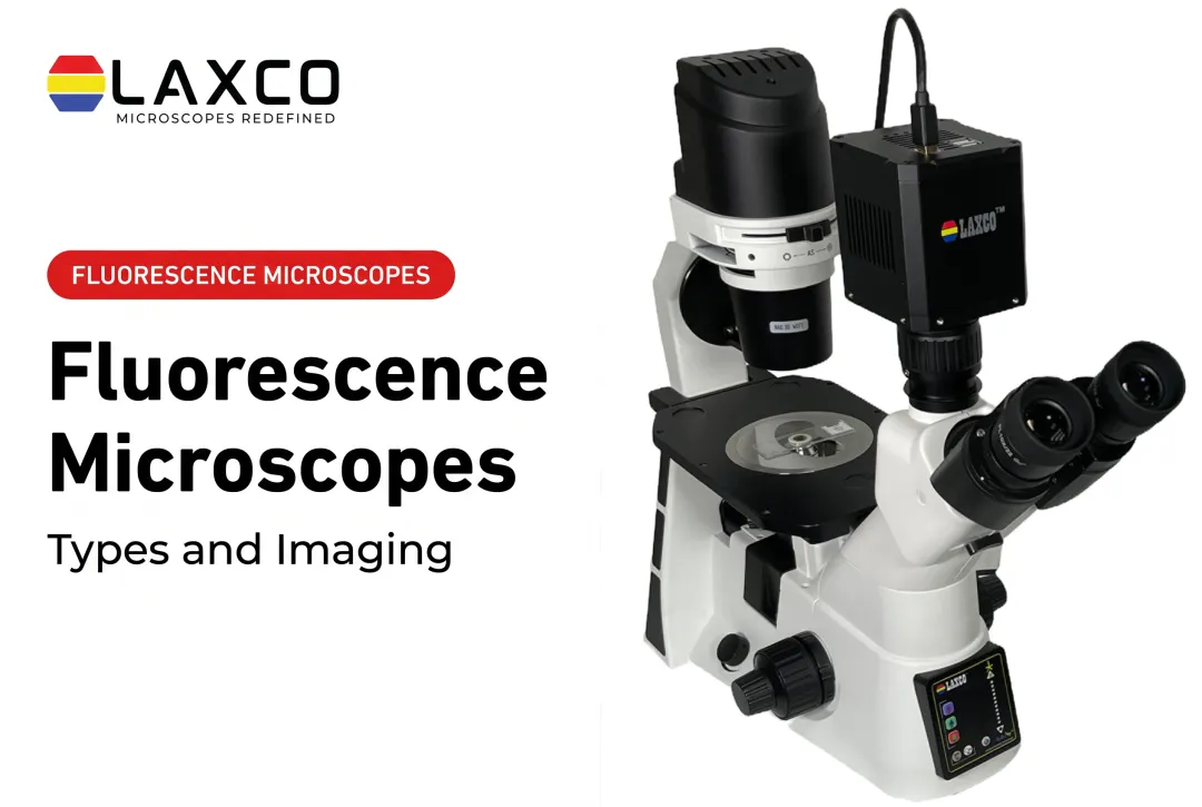
- SLi3Pro: inverted widefield fluorescence microscope with SeBaLIT LED-based illumination. Supports three-color multiplex fluorescence with commonly used DAPI, FITC, and TRITC fluorochromes. It is not a laser-scanning confocal system.
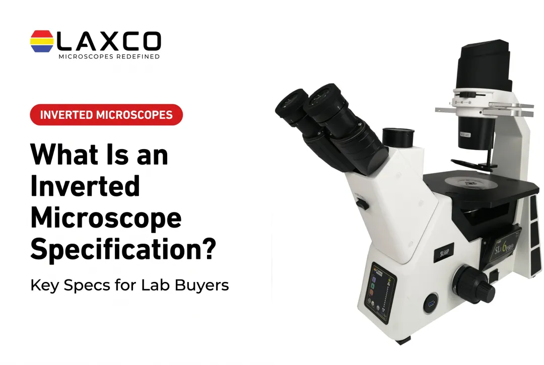
- SLi6Pro: inverted widefield fluorescence microscope with SeBaLIT LED-based illumination covering approximately 390-662 nm and supporting more than 110 fluorochromes. Compatibility with 110+ dyes means spectral fit across many fluorochromes, not 110 simultaneous detection channels. It is not a laser-scanning confocal system.
- Accuva Cellect: a laser capture microdissection system with an infrared laser for capture of single cells or small numbers of cells and a UV laser for cutting dense structures.
- LEAP AI: combines Accuva LEAP optics with Media Cybernetics Image-Pro AI for deep-learning segmentation, measurement, and reporting.
- CellLive: CellLive CL11 and CL22 systems support continuous live-cell imaging inside CO2 incubators, with scheduled time-lapse imaging and remote monitoring for incubator-based observation workflows.
Training, application assistance, and service arrangements should be confirmed with Laxco for the specific instrument and configuration. Researchers comparing fluorescence systems can discuss their imaging requirements and available support directly with a Laxco specialist.
Frequently Asked Questions
When do I need laser illumination rather than LED illumination?
Laser illumination is typically required for point-scanning confocal imaging and some specialized methods that need a focused, narrow-band source. Many routine widefield multichannel fluorescence applications are well served by LED illumination, often with simpler day-to-day operating requirements. The choice depends on the imaging mode, sample thickness, fluorophore panel, and whether optical sectioning is required.
Are the SLi3Pro and SLi6Pro laser microscopes?
No. Both use SeBaLIT LED-based illumination for widefield multicolor fluorescence on an inverted platform. They are not laser-scanning confocal systems. The SLi3Pro supports three-color multiplex fluorescence with commonly used DAPI, FITC, and TRITC fluorochromes. The SLi6Pro covers approximately 390-662 nm with more than 110 compatible fluorochromes; that figure refers to dye compatibility, not simultaneous channel count.
Which Laxco instrument uses lasers?
Accuva Cellect uses an infrared laser and a UV laser for tissue isolation in laser capture microdissection. Those lasers support capture and cutting workflows rather than point-scanning confocal fluorescence imaging. Other Laxco fluorescence systems such as the SLiPro models rely on LED-based illumination for widefield imaging.
What safety documentation should I request for a laser system?
Request the manufacturer's laser classification documents and confirm requirements with your institution's Laser Safety Officer. In the United States, ANSI Z136.1 addresses laser hazard controls, and IEC 60825-1 covers laser product classification. Additional reference material is available in OSHA laser standards and related guidance.
How should I compare quotes for fluorescence systems?
Compare illumination channels, filter sets, objectives, camera, software, service, and training as separate line items. Bring a list of your fluorophores and sample types so a Laxco specialist can recommend a configuration.