Key Takeaways
- Formalin fixation fragments RNA, creates RNA-protein crosslinks, and chemically modifies bases.
- FFPE RNA extraction commonly uses tissue digestion and crosslink-reversal steps, with genomic DNA removal when required. Some sequencing-based expression-profiling methods instead work directly from tissue lysates.
- 3′ mRNA-seq, ribodepletion-based whole-transcriptome sequencing, and probe-based capture suit different research goals.
- DV200 is commonly preferred to RIN for assessing FFPE RNA quality.
- Heterogeneous tissue sections can dilute the signal from a target cell population; laser capture microdissection can isolate that population before extraction.
What Is FFPE Tissue?
FFPE stands for formalin-fixed, paraffin-embedded. Tissue is fixed in formalin to preserve morphology, then embedded in paraffin wax for storage at room temperature. A 2020 review in the Journal of Histochemistry & Cytochemistry described FFPE preservation as a widely used standard in diagnostic pathology and discussed its value for retrospective molecular research.
FFPE archives are useful for RNA-seq research for several reasons:
- Room-temperature storage is less demanding than maintaining frozen archives.
- Archived samples often have associated histology and clinical annotation.
- They support retrospective cohort studies that would take years to assemble prospectively.
How FFPE Processing Affects RNA
Crosslinking and Chemical Modification
Formalin fixation can fragment RNA, create RNA-protein crosslinks, and chemically modify nucleotide bases. These effects complicate extraction and downstream enzymatic reactions, potentially reducing library complexity and increasing technical variability. In a study of FFPE RNA demodification, Wehmas et al. examined how formalin-related damage affected RNA recovery and sequencing quality. Earlier, Masuda et al. (1999) demonstrated methylol additions on all four RNA bases, with modification rates differing among bases.
Depending on the tissue, fixation conditions, RNA recovery, and library-preparation method, FFPE-derived sequencing data may show:
- Residual ribosomal RNA: increased rRNA representation when depletion is less effective.
- Sequencing and mapping artifacts: changes in error rates or the proportion of reads aligning to different genomic regions.
- Coverage bias: uneven transcript coverage, including increased 3′ bias in some workflows.
- Reduced library complexity: higher duplication rates and fewer informative molecules when the usable RNA input is limited.
These effects are not inevitable in every FFPE dataset. Appropriate extraction, library preparation, and quality-control methods can mitigate some of them.

Loss of Poly(A) Tails
RNA fragmentation can separate transcript regions from their poly(A) tails, making poly(A)-dependent enrichment less effective for some degraded FFPE samples. Random-primed total-RNA sequencing with suitable ribosomal RNA depletion is often used when more extensive transcript coverage is needed from fragmented RNA.
In a published protocol for degraded FFPE RNA, Levin et al. (PMC8815082) recommended random-primed total-RNA preparation for highly degraded samples, including those with DV200 below 30%. The authors also showed that DV100, which measures the proportion of fragments longer than 100 nucleotides, can be more informative than DV200 for severely fragmented samples.
These recommendations apply to the workflow and sample conditions evaluated. A DV200 below 30% does not automatically make every poly(A)-dependent method unusable; some validated 3′ RNA-seq protocols can still produce useful expression data from highly degraded material.
Fixation and Storage Conditions
RNA quality depends on fixation time, tissue thickness, and storage conditions, not only block age. Formalin penetrates tissue slowly, so the center of thick specimens may begin to degrade before fixation is complete. FFPE extraction protocols therefore include heat-based crosslink reversal and DNase treatment before library preparation.
RNA-Seq Approaches for FFPE Samples
The appropriate method depends on the research question and the degree of RNA degradation.
3′ mRNA-Seq
3′ mRNA-seq focuses sequencing coverage near the polyadenylated ends of transcripts. Many protocols use unique molecular identifiers (UMIs) to distinguish captured RNA molecules and reduce the effects of PCR duplication. They do not necessarily produce only one sequencing read per transcript. By concentrating reads near transcript ends, these workflows can support cost-efficient gene-expression quantification, although their suitability depends on RNA quality and assay design.
In a 2021 study, Jang et al. (PMC8543821) evaluated UMI-based 3′ mRNA-seq using RNA of different quality and input amounts, including highly degraded FFPE samples. Their reported mapping and correlation results varied with the sample type and protocol. For example, mapping rates of 87–99% and correlations above 0.94 were observed in the evaluated reference-RNA protocol comparisons, while more degraded FFPE samples showed input-dependent results.
The approach is useful for gene-level differential expression analysis when the assay is compatible with the available RNA. Because coverage is concentrated at transcript ends, it is generally less informative than suitable full-length methods for comprehensive isoform or splice-variant analysis.
Whole-Transcriptome Sequencing with Ribodepletion
Random priming combined with ribosomal RNA depletion supports broader transcriptome coverage than methods restricted to polyadenylated RNA. Depending on the protocol, it can detect coding transcripts and non-polyadenylated RNA species, including some long non-coding RNAs. However, reliable isoform and fusion analysis still depends on RNA fragment length, library design, sequencing parameters, and assay validation.
In a 2020 Scientific Reports study, Newton et al. evaluated ribodepletion-based RNA sequencing using more than 3,200 FFPE slide samples. The researchers reported that 86% of extracted RNA samples could proceed through library preparation. After additional quality requirements were applied, the overall RNA-sequencing success rate was 81%.
These figures describe the study's workflow and sample cohort rather than guaranteed success rates for FFPE sequencing generally.
Probe-Based Hybridization Capture
Probe-based hybridization capture uses oligonucleotide probes to enrich selected transcript-derived sequences before sequencing. Depending on the assay, probes may target a defined gene panel or a broader set of coding exons. This approach can improve the representation of relevant transcripts in degraded FFPE material, although coverage depends on probe design and target selection.
A 2023 study comparing exome-capture RNA-seq methods (PMC10722801) evaluated capture-based protocols for degraded RNA and FFPE samples. It highlighted the importance of probe selection, library preparation, sequencing parameters, and downstream analysis when evaluating performance.
Ligation-based expression profiling is a separate approach. In a 2019 study, Trejo et al. (PMC6386473) used TempO-Seq, a targeted, ligation-based whole-transcriptome expression-profiling method, to analyze FFPE tissue through direct tissue lysis. Their workflow did not require conventional RNA purification or reverse transcription. The study reported strong expression agreement between appropriately compared fixed and unfixed samples.
TempO-Seq should therefore not be classified as conventional hybridization-capture RNA-seq. Its results represent expression measurements from the genes covered by the assay's probe design.
Choosing Between Methods
| Research goal | Approach to consider |
|---|---|
| Gene-level differential expression from suitable polyadenylated RNA | 3′ mRNA-seq, with input and degradation tolerance confirmed for the selected protocol |
| Broad coding and noncoding transcript coverage | Random-primed, ribodepletion-based total-RNA sequencing |
| Fusion or isoform investigation | A validated sequencing method with appropriate transcript coverage, read structure, and sensitivity for the available RNA quality |
| Enrichment of selected genes or coding exons from degraded RNA | Probe-based hybridization capture, using a panel or broader exome-capture design |
| Sequencing-based expression profiling directly from small FFPE tissue areas | A compatible direct-lysate, ligation-based method such as TempO-Seq |
Quality Assessment and Input Considerations
DV200 Versus RIN
The RNA Integrity Number (RIN) uses electrophoretic features of ribosomal RNA to assess integrity, but these features are often poorly preserved in fragmented FFPE samples. DV200 measures the percentage of RNA fragments longer than 200 nucleotides and can provide more useful information about the size distribution of degraded RNA.
In a 2020 study by Matsubara and colleagues (PMC7063185), researchers evaluated 71 RNA samples: 30 FFPE tissue samples, 25 fresh-frozen samples, and 16 cell-line samples. Across the combined study population, DV200 correlated more strongly with initial library-product yield (R² = 0.82) than RIN-equivalent measurements (R² = 0.69). These values describe the studied sample set and should not be interpreted as correlations measured solely across FFPE specimens.
DV200 acceptance thresholds depend on the library-preparation chemistry. For severely degraded samples, DV100 may provide additional information. Researchers should follow the chosen assay's quality-control criteria rather than use one universal FFPE RNA threshold.

Input, Depletion, and Sequencing Depth
- Input: some library-preparation methods benefit from higher RNA input when samples are highly degraded, but the requirement depends on the assay and available material. Low-input methods may be appropriate when validated, especially for microdissected samples where additional RNA cannot be obtained.
- Ribosomal depletion: the depletion strategy affects residual rRNA and the retention of low-abundance transcripts.
- Depth: targeted panels generally need less sequencing depth than whole-transcriptome libraries.
- Multiplexing: pooling low-complexity libraries deeply can increase duplicate reads.
Isolating Target Cells from FFPE Sections
FFPE tumor sections often contain tumor cells, stroma, immune infiltrate, and normal tissue. Bulk extraction combines RNA from all these populations, which can dilute signals from the population of interest.
Laser capture microdissection (LCM) isolates selected cells or tissue regions under microscopic guidance before downstream molecular analysis. This allows researchers to enrich defined cell populations instead of averaging RNA from an entire heterogeneous section.
Laser capture microdissection can help researchers distinguish the RNA-expression profiles of cell populations that occupy different regions of a heterogeneous tissue section. Rather than extracting RNA from the entire specimen, researchers can select morphologically or marker-defined cell populations for separate downstream analysis.
A review of RNA-sequencing approaches in clinical oncology describes LCM-assisted RNA-seq as an approach for isolating selected material before transcriptomic analysis. It also discusses the technical challenges of obtaining sufficient RNA quantity and quality from FFPE specimens.
This approach can help reduce the mixing of expression signals from different tissue compartments, although the specificity of the resulting data depends on target selection, capture purity, sample preservation, and downstream RNA-seq methods.
In heterogeneous tumor specimens, the same principle can be applied to separately selected tumor, stromal, vascular, or other histologically identifiable compartments. Comparing RNA profiles from these regions may reveal differences that would be obscured when the entire tissue section is analyzed as one bulk sample.
LCM-assisted transcriptomics is not automatically single-cell transcriptomics. When several cells are captured and analyzed together, the RNA-seq results represent the combined expression profile of the collected population or region. The spatial information comes from the documented location and selection of the captured material, not from a continuous gene-expression map of the entire section.
LCM often provides limited RNA input, so extraction and library preparation must be compatible with the sample amount and RNA quality. Pilot captures can help determine the collection size and processing method required for the study.

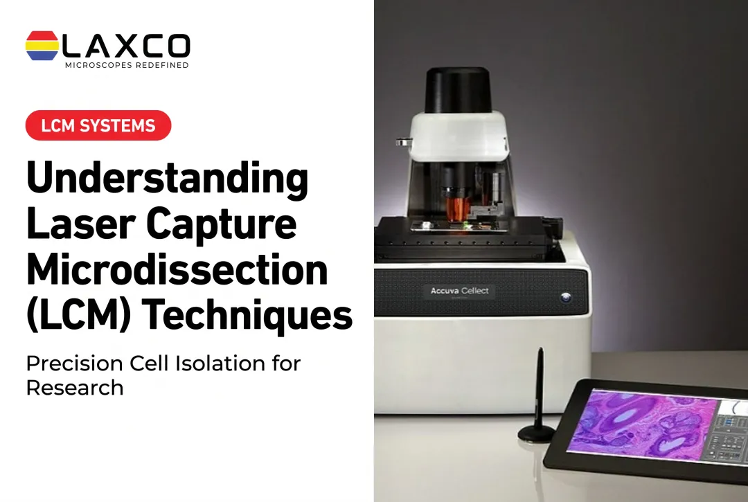
Laxco's Accuva Cellect Laser Capture Microdissection System supports the microscopy-guided identification, selection, and collection of target cells or tissue regions. It forms the tissue-isolation stage of a broader microgenomics workflow, while nucleic-acid purification, amplification, and sequencing take place through separate downstream laboratory procedures. Its documented capabilities include:
- An infrared (IR) laser designed for film capture of single cells or small numbers of cells.
- An ultraviolet (UV) laser designed for cutting dense structures, such as bone or plant tissue.
- A five-step software workflow with visual quality control and documentation.
- Documented support for FFPE and fresh-frozen samples, H&E-stained and LCM-optimized stains, immunofluorescence, unstained samples, and cell culture.
Accuva Cellect isolates the target cells. RNA extraction, library preparation, and sequencing are separate downstream steps performed by the researcher's laboratory.
Frequently Asked Questions
Which sample types can Accuva Cellect isolate for FFPE RNA-seq workflows?
Laxco lists FFPE and fresh-frozen tissue, along with H&E-stained and LCM-optimized stained sections, immunofluorescence, unstained samples, and cell culture. Confirm compatibility with your RNA extraction and library kit during planning.
How many cells should I capture for FFPE RNA-seq?
The number depends on RNA quality, the extraction method, and the library kit's input requirements. Pilot captures with QC measurements help establish the required tissue area before processing a full cohort.
What support does Laxco provide after purchase?
Laxco lists training and 48-hour response times for Accuva Cellect. Confirm which training and support formats apply to your configuration. On-site, virtual, or live delivery should not be assumed unless specified in your agreement.
How much bench space does Accuva Cellect require?
Accuva Cellect is described as about 32% smaller than comparable systems; confirm current dimensions with Laxco. Allow additional room for control equipment, safe operation, slide handling, and consumables. See the Accuva Cellect product page.