Key Takeaways
- Organoids retain tissue architecture and expression features that 2D cultures often lose.
- Published studies report promising but imperfect agreement between organoid and patient drug responses.
- Heterogeneity, manual handling, and imaging complexity limit reproducibility.
- Automated imaging and validated image analysis support measurements at the single-organoid level.
What Is Organoid Screening?
Organoids are 3D structures grown from adult stem cells, induced pluripotent stem cells (iPSCs), or tumor tissue. They self-organize into structures that resemble features of the source organ.
A representative organoid screening workflow (preparation methods and timelines vary by model and assay):
- Isolate cells from a biopsy, stem-cell source, or tumor tissue.
- Embed the cells in an extracellular matrix.
- Culture the structures until they mature, often over several weeks.
- Expose the organoids to test compounds.
- Measure viability, growth, and morphological changes.

Research Applications
- Oncology drug screening using tumor-derived organoids (tumoroids).
- Toxicity testing of compounds at the preclinical stage.
- Patient-derived models for studying variation in drug response between donors.
- Disease modeling with cerebral, lung, and intestinal organoids.
A 2021 pooled analysis reported 81% sensitivity and 74% specificity for patient-derived organoids predicting treatment response in the studies reviewed (npj Precision Oncology, 2021). The authors noted a risk of selection bias across those studies. These results support organoids as research models rather than stand-alone predictors.
Why Organoids Are Used in Drug Discovery
Compared with Traditional Models
Organoids can preserve histological features and gene-expression patterns of the source tissue. Compared with animal studies, they:
- Raise fewer ethical concerns for large-scale screening.
- Can reduce long-term cost once protocols are standardized.
- Typically produce results faster than in vivo experiments.
Translational Relevance
Many drug candidates fail in clinical development, and limited translatability of preclinical models is one contributing factor. Human-derived organoids are used to add human tissue context earlier in the pipeline. They do not reproduce systemic effects such as metabolism, vascular supply, or a full immune system.
Technical Challenges in Organoid Screening
- Population heterogeneity: organoids in the same culture vary in size, growth rate, and viability. Pooled readouts average these differences.
- Manual handling: matrix seeding is labor-intensive, and well-to-well variability increases when results depend on individual technique.
- Imaging complexity: organoids are 3D, overlap, and vary in shape. Multifocal imaging is often required, and data volumes make tracking individual organoids difficult.
- Limited standardization: protocols differ between labs, which complicates cross-study comparison.

Pooled endpoints can hide outlier organoids that respond differently to treatment. Single-organoid measurements retain this information.
Designing a Reproducible Organoid Drug Screen
Reliable organoid screening requires more than growing 3D cultures and exposing them to compounds. Differences in organoid size, cell composition, passage history, extracellular matrix, plating density, and imaging conditions can influence measured drug responses.
Before scaling a screen, laboratories should establish a reproducible assay with appropriate controls and predefined analysis criteria.
Step 1 — Standardize the Starting Organoids
- Define the organoid model. Record the tissue source, culture conditions, passage information, and relevant biological characteristics.
- Control starting material. Establish appropriate organoid number, size distribution, or cell input per well according to the assay.
- Document matrix conditions. Record extracellular matrix type, preparation, and batch information when these may influence growth or compound exposure.
- Assess baseline viability. Confirm that organoids are healthy and sufficiently consistent before treatment.
Starting conditions should be standardized within the study rather than assumed to be identical across different organoid models.
Step 2 — Design Controls and Replication
Include controls capable of distinguishing compound effects from ordinary culture variability.
- Vehicle controls: account for the solvent or carrier used to deliver the compounds.
- Positive-response controls: confirm that the assay detects the intended type of biological response.
- Background controls: measure assay signals unrelated to viable organoids where relevant.
- Technical replicates: help evaluate variation between wells within the same experiment.
- Biological replicates: help establish whether findings persist across independent cultures, preparations, or model sources.
Distribute controls appropriately across the plate and consider edge effects, evaporation, and dispensing-related variation.
The number of replicates should be planned according to the assay's variability, the intended effect size, and available material.
Step 3 — Define Compounds, Exposure, and Endpoints
Before testing, establish:
- The compound concentration range and appropriate vehicle concentration.
- The exposure duration and imaging or assay time points.
- The primary endpoint, such as viability, growth inhibition, morphology, or a defined molecular response.
- Any secondary measurements used to distinguish biological effects from image-analysis artifacts.
- The criteria for excluding unsuitable wells or failed measurements.
For example, a reduction in projected organoid size may indicate slower growth or structural changes but does not independently prove cell death. Viability conclusions require a suitable, validated measurement.
Step 4 — Evaluate Plate Quality
Assess the consistency of controls and measurement performance before interpreting treatment effects.
Useful screening-quality measures may include:
- Control-well coefficient of variation.
- Signal-to-background or signal-to-noise ratio.
- Assay dynamic range.
- Z′ factor, where suitable positive and negative controls are available.
- Agreement between independent experimental runs.
The Z′ factor evaluates control separation relative to variability. Although values above 0.5 are commonly considered favorable in conventional high-throughput screening, no single threshold establishes that every organoid assay is valid.
Complex organoid assays may require additional quality criteria and assay-specific acceptance limits.
A review of organoid-based image screening discusses controls, replication, imaging endpoints, and sources of experimental variability. A published cancer organoid drug-sensitivity protocol provides further examples of plate-quality assessment and downstream analysis.
Step 5 — Validate Hits and Biological Interpretation
Initial screening results should be treated as candidate findings rather than confirmed biological effects.
For promising compounds:
- Repeat the experiment using independent preparations where feasible.
- Perform concentration-response analysis to evaluate the consistency of the effect.
- Confirm the result using a complementary measurement where appropriate.
- Review image segmentation and excluded wells for systematic errors.
- Compare results across relevant organoid models or donor-derived samples when the research question requires it.
Single-organoid measurements can reveal differences hidden by pooled well-level assays. However, apparent resistant or sensitive subpopulations should be supported by suitable biological and statistical evidence.
Where LAXCO Fits
For image-based screening, repeatable acquisition and consistent quantitative analysis are important.
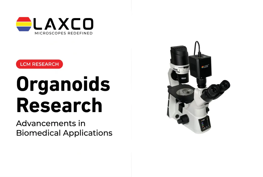
Laxco's LEAP AI combines Accuva LEAP microscopy with Image-Pro AI software for segmentation, measurement, and reporting. The analysis workflow should be validated for the relevant organoid type, imaging modality, and experimental endpoint.
For longer-term monitoring, CellLive provides incubator-compatible imaging with scheduled acquisition. Available imaging modalities depend on the selected CellLive model.
These systems support defined imaging and image-analysis steps. They do not independently perform compound dispensing, organoid culture preparation, or complete drug-screening validation.
Imaging and Image Analysis for Organoid Screens
Automated imaging allows individual organoids to be followed over time instead of measured at a single pooled endpoint. Deep-learning segmentation applies the same criteria across images once validated for the organoid type. Batch processing reduces manual analysis and improves consistency across plates.
Laxco's LEAP AI combines Accuva LEAP optics with Media Cybernetics Image-Pro AI. It follows an Image → Process → Measure → Report workflow.

Its capabilities include:
- Pre-trained deep-learning models for AI-assisted segmentation.
- Batch-processable life-science protocols with no-code options.
- Real-time data visualization.
- Customizable reporting.
For longer time courses, Laxco's CellLive systems support scheduled image acquisition inside CO₂ incubators without repeatedly removing cultures from their controlled environment.
CellLive CL11 provides brightfield time-lapse monitoring. CellLive CL22 adds phase contrast, multichannel fluorescence, and Z-stack scanning for experiments requiring additional imaging capabilities.
The imaging configuration must match the biological endpoint. Automated segmentation and measurement should be validated against representative manually reviewed images, particularly when organoids overlap or when measurements depend on depth-resolved structures.
Cost Considerations
Organoid screening costs come from several sources:
- Culture reagents, including extracellular matrices that can vary between batches.
- Imaging equipment suited to 3D, multifocal data.
- Skilled labor for seeding, feeding, and quality control.
- Assay validation time, which increases with panel size.
In-house screening requires investment in equipment and training but builds internal expertise. Outsourcing converts capital costs into per-study fees. When comparing contract research quotes, confirm what each includes, such as model generation, imaging, raw data delivery, and repeat criteria.
Scale also matters. A single-model screen requires fewer plates, less imaging time, and less data storage than a multi-model panel.
Frequently Asked Questions
Which Laxco system supports organoid image analysis?
LEAP AI combines Accuva LEAP optics with Image-Pro AI for AI-assisted segmentation, measurement, and reporting. Batch protocols allow consistent analysis across plates, subject to validation for your organoid type.
Can organoids be imaged without leaving the incubator?
Laxco's CellLive systems are designed for continuous imaging inside CO₂ incubators. They support scheduled time-lapse acquisition, which suits tracking organoid growth over several days.
How long does it take to grow organoids for screening?
The timeline depends on whether researchers are establishing a new organoid model or preparing an existing culture for testing.
Establishing and expanding a new organoid line may require several weeks or longer, depending on the tissue source and culture protocol. Established organoid cultures can often be replated, allowed to recover or grow, and exposed to compounds over shorter assay-specific periods.
The appropriate timeline depends on organoid growth characteristics, assay format, treatment design, and the biological endpoint. Laboratories should establish consistent starting conditions and validate the screening schedule for their model.
How do organoids compare with patient-derived xenograft (PDX) models?
Organoids are in vitro models that are generally easier to scale for parallel compound testing and may offer faster experimental turnaround once suitable cultures are established.
Patient-derived xenografts (PDXs) grow in animal hosts, typically immunodeficient mice. They provide an in vivo environment that includes host-derived vasculature, systemic drug exposure, and interactions with surrounding tissues.
However, PDX models do not recreate a fully human tumor microenvironment. Human stromal components are often replaced by mouse-derived stroma during passaging, and the use of immunodeficient hosts limits the study of normal immune responses.
Organoids and PDX models therefore offer complementary advantages and limitations. The preferred model depends on whether the study prioritizes scalable compound testing, tissue-specific biological responses, host interactions, or in vivo drug effects.
The NCI PDXNet consensus recommendations discuss these strengths and limitations.