Key Takeaways
- Cytology examines individual cells, so resolution, illumination, and stain compatibility are central requirements.
- Brightfield, phase contrast, fluorescence, and digital systems suit different sample types and workflows.
- Numerical aperture and Köhler alignment matter more than magnification alone.
- AI-assisted analysis requires validation for each sample type and intended use.
What Is Cytology?
Cytology is the microscopic study of individual cells, as distinct from histology's focus on tissue architecture (NCBI Bookshelf). Samples are typically less invasive to collect than surgical biopsies.
Common collection methods:
- Fine needle aspiration (FNA) from masses or lesions.
- Fluid preparations from pleural, peritoneal, or cerebrospinal samples.
- Touch imprints made by pressing a tissue core onto a slide.
- Scrapings and brushings from mucosal surfaces.
Samples are usually stained before viewing. Wet-fixed smears commonly receive the Papanicolaou stain. Air-dried preparations use Romanowsky-type stains, which highlight cytoplasmic detail (PMC, 2016).
Why Optical Quality Matters
Fine nuclear detail, such as chromatin pattern and nuclear membrane contour, requires adequate resolution and even illumination. Poor optics or misaligned illumination reduce the visibility of this detail and slow slide review.
Types of Microscopes Used in Cytology
| Method or configuration | Commonly used for | Example use |
|---|---|---|
| Brightfield | Stained slides | Papanicolaou- and Romanowsky-stained smears |
| Phase contrast | Unstained, translucent samples | Live or minimally processed research samples |
| Fluorescence | Labeled cellular components | Marker studies in research |
| Digital, all-in-one | Shared viewing and teaching | Group review and image documentation |
Digital systems are an imaging and documentation configuration, not a distinct optical contrast method.
Brightfield microscopes transmit light through the stained specimen and are the standard format for reviewing stained smears (PMC). Laxco's LMC-4000 Series upright microscopes support brightfield, darkfield, phase contrast, and fluorescence methods, with infinity-corrected optics and full Köhler illumination. Configuration should match the sample and workflow.
Phase contrast microscopes convert small phase shifts in transparent specimens into visible contrast. This allows unstained cells to be examined without fixation. Laxco's LMI3000 is an inverted microscope used for phase contrast observation of cultured cells.
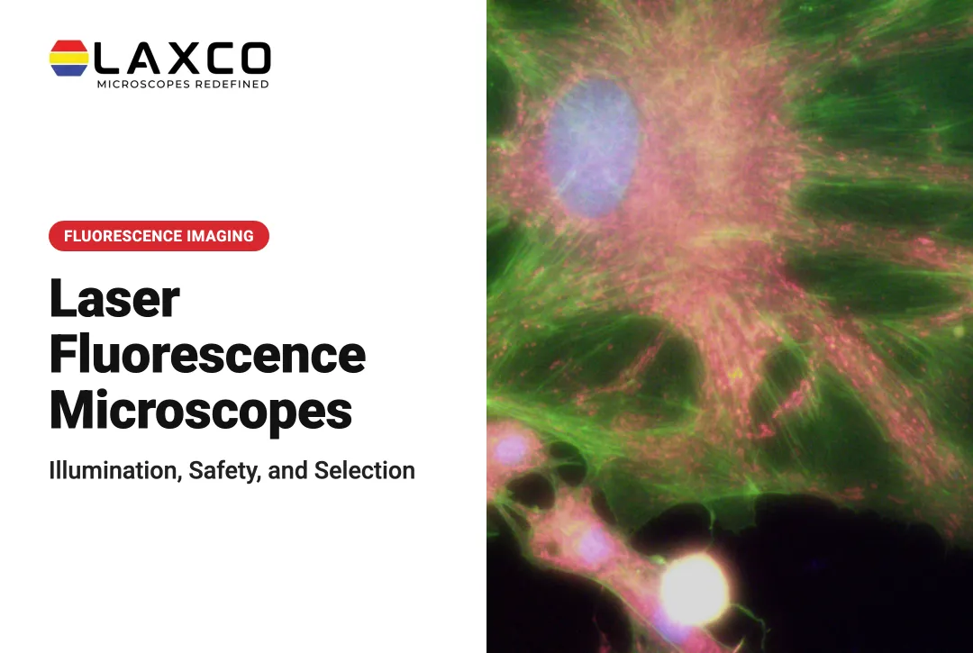
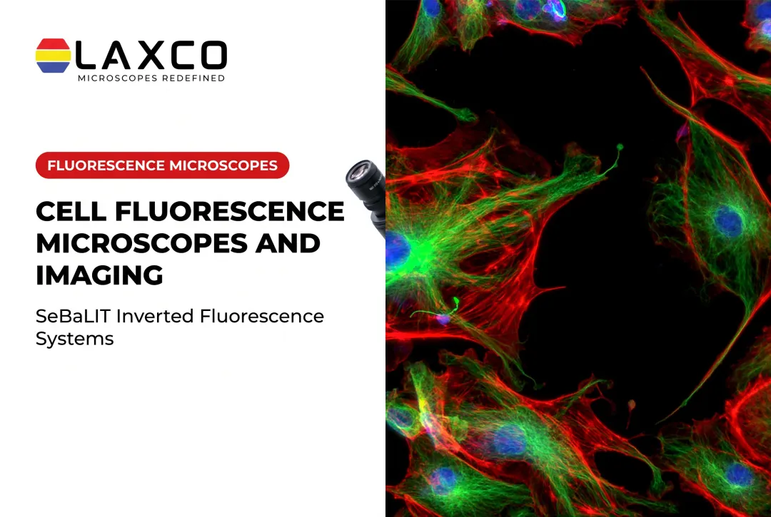
Fluorescence microscopes excite labeled cells at specific wavelengths and separate the emitted signal from background light:
- The SLi3Pro supports common DAPI, FITC, and TRITC fluorochromes.
- The SLi6Pro is an inverted widefield fluorescence microscope with SeBaLIT™ illumination that supports three-color multiplex fluorescence across approximately 390–662 nm and lists compatibility with more than 110 fluorochromes. That figure describes fluorochrome compatibility, not simultaneous imaging of more than 110 markers.
Digital All-in-One Systems
Digital microscopes combine the camera, computer, and display in one unit. Laxco's SeBa2 provides touchscreen viewing, HDMI projection for group review, and image saving to USB or the SeBaShare cloud. These features support teaching, collaboration, and image documentation.
Laser Capture Microdissection
Some research studies link cell morphology to molecular analysis. Laser capture microdissection (LCM) isolates selected cells or regions under direct visualization for downstream DNA, RNA, or protein work (PMC). LCM is a research isolation tool, not a substitute for routine cytology review, and it is not universally validated for every smear type.
Laxco's Accuva Cellect Laser Capture Microdissection System uses an infrared (IR) laser designed for film capture of single cells or small numbers of cells. A UV laser is designed for cutting dense structures. Documented sample types include FFPE and fresh-frozen tissue, stained and unstained sections, immunofluorescence, and cell culture.
Features to Evaluate in a Cytology Microscope
Objectives and Resolution
For routine examination of stained cytology slides, an upright compound microscope with appropriate 10×, 40×, and 100× oil-immersion objectives provides flexibility for screening and examining fine cellular and nuclear details. Resolution depends primarily on numerical aperture rather than magnification alone, and suitable illumination and specimen preparation are essential for clear images.
Microscope selection should follow the sample type and intended workflow. Laboratories examining conventional Papanicolaou- or Romanowsky-stained smears should generally prioritize upright brightfield optics, Köhler illumination, comfortable slide handling, and suitable high-resolution objectives. Inverted phase contrast microscopes are more appropriate for many live-cell and culture-vessel studies, while inverted fluorescence microscopes support research involving fluorescently labeled cellular markers. Digital microscope configurations can add image documentation and collaboration capabilities but do not replace the underlying optical requirements.
Illumination and Alignment
Köhler illumination provides even field brightness and controlled contrast. A centered condenser with accessible field and aperture diaphragms supports consistent alignment between users. The LMC-3000 and LMC-4000 series include full Köhler illumination.
Camera Integration and Head Configuration
A trinocular head allows a camera to be attached while the eyepieces remain in use, which supports documentation and teaching. Laxco options include:
- LMC-4000 Series with an ergonomic trinocular head.
- SLi3Pro and SLi6Pro fluorescence systems.
- SeBa digital microscopes with an integrated camera and display.

Software
Image-analysis software should support your file formats, measurement needs, and batch processing across slides. Software used for any regulated workflow requires separate validation by the laboratory.
Cytology Compared With Related Disciplines
Cytology overlaps with several related laboratory disciplines, but the samples and analytical objectives differ:
- Cytology and microbiology: cytology focuses on the morphology and characteristics of human or animal cells, while microbiology studies microorganisms such as bacteria and fungi (microbiology reference).
- Cytology and histology: cytology examines individual cells or cell groups from smears, aspirates, and fluid preparations. Histology examines tissue sections while preserving the surrounding tissue architecture.
- Exfoliative and aspiration cytology: exfoliative cytology examines naturally shed or surface-collected cells, while aspiration cytology collects cellular material from a lesion or mass using a needle.
AI-Assisted Imaging in Cytology Research
Research into artificial intelligence for digital cytopathology has expanded substantially. A 2025 narrative review of digital cytopathology identified 151 publications, with 78.1% published during the preceding decade.
Researchers have investigated AI-assisted cell identification, image classification, segmentation, and prioritization of potentially abnormal cells for further review. However, results depend on specimen preparation, imaging conditions, training datasets, model design, and independent validation.
These research applications should not be confused with documented performance or regulatory clearance for LAXCO instruments. AI-assisted image analysis may support research workflows, but use in clinical cytology screening requires evidence and authorization appropriate to the specific system and intended application.

For research image analysis, Laxco's LEAP AI is a research imaging platform, not a cervical screening system. It pairs Accuva LEAP inverted microscope optics with Media Cybernetics Image-Pro AI and uses pre-trained deep-learning models for segmentation and measurement across an Image → Process → Measure → Report workflow.

Its capabilities include:
- Batch-processable life-science protocols with no-code options.
- Pre-trained models intended to reduce variability between users.
- Applications in cell biology, cancer research, developmental biology, and drug discovery.
Laxco's LEAP Grant Assistance Program provides Grant Language Kits for proposals that include Accuva LEAP to agencies such as NIH, NSF, HHMI, and DOD.
Frequently Asked Questions
Which Laxco upright microscope suits multiple contrast methods?
The LMC-4000 Series supports brightfield, darkfield, phase contrast, fluorescence, and metallurgical methods. It includes infinity-corrected optics, an ergonomic trinocular head, and full Köhler illumination.
Which Laxco option suits group review and teaching?
The SeBa2 digital microscope combines camera, computer, and touchscreen display. Images can be projected over HDMI or saved to USB or the SeBaShare cloud for sharing.
What equipment does a cytology workflow need?
Core equipment includes a compound microscope with oil-immersion objectives, slides, stains, and collection tools. Cameras and image-analysis software are common additions for documentation and measurement.
Can bacteria be seen at 1000x magnification?
Yes. A 100x oil-immersion objective with a 10x eyepiece gives about 1000x total magnification, which is commonly used for bacteria (UC Davis). Resolution and staining quality matter as much as magnification.