Key Takeaways
- scRNA-seq resolves cell identity and state within heterogeneous tumor samples.
- The TME includes malignant, immune, stromal, and endothelial cells whose interactions can be studied from expression data.
- Dissociation method, storage, and handling influence data quality and can introduce artifacts.
- Computational calls, such as malignant-cell inference and cell-communication analysis, generate hypotheses that need orthogonal validation.
Why Single-Cell Resolution Matters in Tumor Biology
Tumors are heterogeneous. A single biopsy can contain:
- Malignant cells in different transcriptional states.
- Immune cells in exhausted or activated states.
- Stromal cells with distinct gene-expression programs.
Bulk RNA sequencing produces one averaged value per gene. A 2021 review noted that bulk methods can obscure signals from rare cell populations. Some of these populations are linked to tumor progression and drug resistance (International Journal of Oral Science, 2021).
scRNA-seq uses cell-specific barcoding or other single-cell library methods to generate expression profiles for individual cells. In a 2018 lung tumor atlas, Lambrechts and colleagues profiled 52,698 cells in their discovery dataset and identified extensive diversity among stromal populations, including fibroblasts, endothelial cells, and immune cells. Their findings demonstrated how single-cell analysis can resolve cellular differences that bulk measurements may obscure.

Identifying Malignant Cells
Distinguishing malignant from non-malignant cells is a recurring analytical challenge. Tools such as InferCNV and CopyKAT infer copy-number variation from expression data to flag likely malignant cells. Benchmarking studies have reported variable accuracy across tumor types. Near-diploid tumors are particularly difficult to classify using copy-number inference alone.
What scRNA-Seq Reveals About the Tumor Microenvironment
The TME consists of malignant cells, immune cells, cancer-associated fibroblasts (CAFs), endothelial cells, and extracellular matrix. CAFs remodel the extracellular matrix and can influence immune-cell infiltration. This remodeling contributes to desmoplasia, the dense fibrous stroma found in many solid tumors.

Inferring Cell-Cell Communication
Tools such as CellPhoneDB and CellChat analyze ligand and receptor expression to propose communication between cell types:
- CellPhoneDB identifies ligand-receptor interactions statistically enriched between cell-type pairs.
- CellChat models communication probability using expression data and curated ligand-receptor databases.
- Limitation: co-expression suggests a possible interaction but does not demonstrate that it occurs in tissue.
Rare Cell States
Some cell states relevant to relapse or resistance are present at low frequency. Single-cell studies in leukemia, for example, have described rare stem-like subpopulations associated with relapse. Such findings depend on capturing enough cells to detect rare states.
Spatial Context
Conventional scRNA-seq identifies cell types and transcriptional states but generally loses their original location when tissue is dissociated. Computational approaches can help recover spatial context by combining single-cell profiles with spatial transcriptomics data. For example, CellTrek maps single-cell transcriptomes to tissue coordinates and has been used to investigate the spatial organization of tumor subclones and immune-cell states in breast ductal carcinoma in situ.
From Tissue to Transcriptome: The scRNA-Seq Workflow
A typical tumor scRNA-seq workflow has five stages:
- Tissue collection and preservation: time to processing and storage method affect RNA quality and cell viability.
- Dissociation or nuclei isolation: tissue is separated into single cells or nuclei.
- Cell capture and library preparation: transcripts are barcoded for sequencing.
- Sequencing: libraries are sequenced to generate reads.
- Computational analysis: quality control, clustering, and cell-type annotation.

Dissociation Artifacts
Enzymatic dissociation at 37 °C can induce stress-response and heat-shock genes that were not expressed in the original tissue. Cold-active dissociation protocols and single-nucleus approaches are used to reduce this effect. Cryopreservation can also reduce the proportion of viable cells recovered.
Targeted Isolation from Tissue Sections
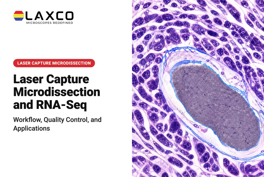
When a study focuses on a defined tumor region, laser capture microdissection (LCM) can isolate selected cells or areas from a tissue section before further processing. The operator selects targets under the microscope, preserving the link between the sample and its histological context. LCM material may be processed as a pooled multi-cell sample or, when yield and chemistry allow, as individual cells for single-cell library preparation. Foley et al. (2019) distinguish these paths clearly: LCM can collect multiple cells of a selected type for bulk or multi-cell analysis, or capture single cells that are then processed with single-cell RNA-seq methods (PMC6836736). Throughput is lower than suspension-based capture, and downstream success depends on RNA input, preservation method, and the chosen library chemistry.
Laxco's Accuva Cellect Laser Capture Microdissection System supports this approach:
- An infrared (IR) laser designed for film capture of single cells or small numbers of cells.
- An ultraviolet (UV) laser designed for cutting dense structures, such as bone or plant tissue.
- A five-step software workflow with visual quality control and documentation.
- Documented support for FFPE and fresh-frozen samples, H&E-stained and LCM-optimized stains, immunofluorescence, unstained samples, and cell culture.
Image-Based Selection of Regions of Interest
Microscopy helps identify regions of interest before capture. LEAP AI is a separate Laxco imaging platform that combines Accuva LEAP optics with Media Cybernetics Image-Pro AI for AI-assisted segmentation and measurement. It is not integrated into Accuva Cellect capture control. Automated segmentation can reduce manual workload, but results should be validated for each tissue type and stain.
Quality Control Checkpoints
- Sample viability and RNA quality before library preparation, matched to the chosen chemistry.
- Cell or nucleus recovery rate after dissociation or isolation, with notes on any enrichment steps.
- Ambient RNA and empty-droplet or empty-well rates after capture.
- Doublet or multiplet rates and mitochondrial-content filters during quality control.
- Batch structure across runs, operators, and sample lots before integrating datasets.
Applications of scRNA-Seq in Cancer Research
Tumor Classification and Biomarker Research
Large scRNA-seq studies have proposed new ways to classify tumor cell states. In colorectal cancer, Joanito et al. (2022) described intrinsic consensus molecular subtypes (iCMS2 and iCMS3) of malignant epithelial cells (Nature Genetics). In breast cancer, Wu et al. (2021) described recurring cellular ecotypes associated with differences in clinical outcome (Nature Genetics). These associations come from the studied cohorts and require further validation.
Immunotherapy Response
Longitudinal scRNA-seq studies in melanoma have compared patients who respond to immunotherapy with those who develop resistance. Such studies have associated specific tumor subclones and immune-cell states with response or resistance.
Tumor Evolution and Drug-Discovery Research
Sampling tissue across disease stages allows researchers to reconstruct transcriptional changes during progression. In drug-discovery research, scRNA-seq can help assess whether cell lines or organoids reflect the heterogeneity of the source tumor.
Limitations of scRNA-Seq
- Technical noise: dropout, batch effects, and dissociation artifacts complicate clustering and annotation.
- Malignant-cell inference: copy-number inference tools vary in accuracy, particularly for near-diploid tumors.
- Cost: per-cell library preparation and sequencing make scRNA-seq more expensive than bulk RNA-seq.
- Sampling: capturing more cells improves detection of rare states but does not correct biased sampling or poor viability.
Findings from scRNA-seq should be validated using orthogonal methods, such as immunohistochemistry, spatial profiling, or functional assays.
Frequently Asked Questions
Should I dissociate tumor tissue or use single-nucleus sequencing?
Single-nucleus sequencing is often used for frozen tissue or tissues that dissociate poorly, and it can reduce dissociation-induced stress signatures. Whole-cell sequencing captures cytoplasmic transcripts. The choice depends on sample storage, tissue type, and the transcripts of interest.
How is scRNA-seq different from spatial transcriptomics?
scRNA-seq profiles dissociated cells and loses tissue location. Spatial transcriptomics measures expression in place, although some methods combine signals from several cells per measurement area. The two are often integrated computationally.
When is laser capture microdissection useful in tumor studies?
LCM is useful when molecular data must come from histologically defined regions or cell populations, such as a tumor margin or an immune aggregate. Material can feed pooled multi-cell libraries or, when yield allows, single-cell library preparation. Accuva Cellect supports IR film capture and UV cutting on FFPE and fresh-frozen sections for these workflows.
Which sample types does Accuva Cellect support?
Laxco lists FFPE and fresh-frozen tissue, H&E-stained and LCM-optimized stained sections, immunofluorescence, unstained samples, and cell culture. Confirm compatibility with your RNA workflow during planning.