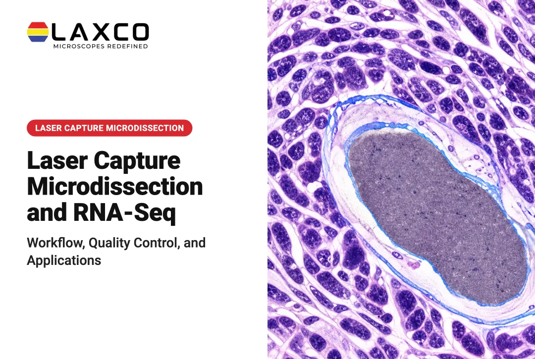
For tissue-based research, preparation begins even earlier: selecting which cells or tissue regions should contribute to the measurement. Homogenizing an entire specimen can dilute signals from the cell population of interest.
This guide compares common preparation methods, provides representative workflows for plasma small-molecule analysis and bottom-up proteomics, explains quality-control requirements, and shows where targeted tissue isolation using laser capture microdissection can support downstream analysis.
Key Takeaways
- Matrix components such as proteins, lipids, and salts can cause ion suppression and instrument contamination.
- No single protocol suits every analyte; the method depends on analyte chemistry, matrix, and required sensitivity.
- Most workflows involve extraction, cleanup, digestion or derivatization, concentration, and introduction into the instrument.
- For tissue studies, sample composition affects what the MS result represents.
Why Sample Preparation Affects MS Results
Sample preparation serves three main purposes:
- Protecting the chromatography column and ion source from particulates and proteins.
- Improving chromatographic separation.
- Increasing analyte signal relative to background.
Inadequate preparation can produce matrix effects. These are changes in analyte response caused by co-eluting compounds such as salts, lipids, and phospholipids. They appear as ion suppression or enhancement during ionization. Tandem MS does not remove this problem, because suppression occurs before fragmentation (LCGC review).
For regulated bioanalytical assays supporting drug development and regulatory submissions, the ICH M10 bioanalytical method validation guideline provides specific requirements for evaluating matrix effects. For applicable chromatographic assays, it generally calls for testing low- and high-concentration quality controls across at least six independent matrix sources or lots, with defined accuracy and precision acceptance criteria, subject to the guideline's stated exceptions.
These requirements should not be treated as universal validation rules for discovery proteomics, untargeted metabolomics, or every other mass-spectrometry workflow. Research laboratories should establish matrix-effect and quality-control procedures appropriate to the analytical method and intended use.
Residual proteins and nonvolatile salts can also contaminate the ion source and column. This increases cleaning frequency and instrument downtime.
Core Sample Preparation Techniques
The appropriate method depends on the matrix, the analyte's chemistry, and the degree of cleanup required.
Dilution ("Dilute and Shoot")
The sample is diluted and injected directly. This approach is used for relatively clean matrices such as urine or cerebrospinal fluid.
- Advantages: fast, with minimal analyte loss.
- Limitations: matrix components remain, which can reduce sensitivity and reproducibility.
Protein Precipitation (PPT)
An organic solvent, such as acetonitrile or methanol, is added to precipitate proteins from serum, plasma, or whole blood. The precipitate is then removed by centrifugation or filtration. PPT is simple and fast, but phospholipids and other lipids often remain in the supernatant.
Liquid-Liquid Extraction (LLE) and Solid-Phase Extraction (SPE)
LLE partitions the analyte between two immiscible liquid phases. Recovery depends on pH, salt content, and solvent choice. SPE passes the sample through a sorbent that selectively retains and then releases the analyte.
- LLE: broad applicability, but very polar compounds may extract poorly.
- SPE: higher selectivity and matrix removal, with more method development and processing time.
Supported Liquid Extraction (SLE) and Phospholipid Removal
SLE absorbs the aqueous sample onto a solid support and extracts it with an immiscible organic solvent. This avoids manual phase separation. Phospholipid-removal plates target phospholipids, which are a major source of matrix effects in blood-derived samples.
Proteomics-Specific Preparation
Protein samples require additional steps before MS analysis:
- Reduction: disulfide bonds are reduced, commonly with dithiothreitol (DTT) or TCEP.
- Alkylation: cysteine residues are alkylated, commonly with iodoacetamide.
- Digestion: proteins are digested into peptides, commonly with trypsin, Lys-C, or both.
Reagent concentrations, denaturants, and digestion times vary by protocol and sample type.
Method Comparison
| Technique | Complexity | Matrix removal | Typical use |
|---|---|---|---|
| Dilute and shoot | Low | Minimal | Relatively clean matrices such as urine |
| Protein precipitation | Low to medium | Moderate | Serum, plasma, whole blood |
| LLE | Medium | Moderate to high | Many small molecules |
| SPE | Medium to high | High | Complex matrices, trace analytes |
| SLE and phospholipid removal | Medium | High for phospholipids | Plate-based bioanalysis |
A General Five-Step Workflow
Most MS preparation workflows follow five stages, although details vary by technique:
- Extraction: isolate the analyte or protein from tissue, serum, or cell lysate.
- Cleanup: remove detergents, salts, and particulates by desalting, filtration, or precipitation.
- Digestion or derivatization: digest proteins enzymatically, or derivatize small molecules to improve ionization or chromatography.
- Concentration and reconstitution: evaporate and reconstitute the sample in an MS-compatible solvent.
- Introduction: inject the sample into the LC-MS or GC-MS system.
The ionization method depends on the analyte. Electrospray ionization (ESI) is a soft method for nonvolatile analytes in solution and can produce multiply charged ions. Electron ionization (EI) is used after gas chromatography for volatile, thermally stable compounds and typically produces singly charged ions.

Representative Mass Spectrometry Sample Preparation Protocols
There is no single protocol suitable for every analyte, tissue, and analytical platform. The following examples show how common preparation methods are organized and where important quality-control decisions occur.
They are representative research workflows, not universal validated standard operating procedures. Reagent quantities, incubation conditions, centrifugation parameters, and instrument settings must be confirmed for the specific method.
Protocol 1 — Plasma Protein Precipitation for Small-Molecule LC-MS/MS
Application: Preparing plasma samples for analysis of suitable small-molecule compounds by LC-MS/MS.
Principle: Organic solvent precipitates plasma proteins, allowing the analyte-containing liquid phase to be separated for further analysis.
- Prepare and identify samples. Label plasma samples, blanks, calibration standards, and quality controls. Add a suitable internal standard at the validated stage of preparation.
- Add precipitation solvent. A commonly studied example is three volumes of acetonitrile to one volume of plasma. The solvent choice and ratio must be validated for the analytes being measured.
- Mix the samples. Mix sufficiently to precipitate proteins, using the validated mixing and temperature conditions.
- Separate precipitated material. Centrifuge according to the method and transfer the clarified supernatant without disturbing the protein pellet.
- Prepare for injection. Where required, dilute, concentrate, or reconstitute the extract in an LC-compatible solvent. Ensure the final solvent composition is appropriate for the chromatography method.
- Evaluate the extract. Check recovery, carryover, matrix effects, and analyte response using matrix-matched controls.
- Proceed to LC-MS/MS. Inject only after the extract and analytical method meet the relevant quality-control criteria.
Critical limitation: Protein precipitation is fast but does not necessarily eliminate phospholipids or other interfering compounds. Additional SPE or phospholipid-removal steps may be necessary for sensitive assays.
Supporting reference: Published plasma protein-precipitation LC-MS/MS method.
Protocol 2 — Bottom-Up Proteomics Sample Preparation for LC-MS/MS
Application: Preparing protein extracts from cells or tissue for peptide-based mass spectrometry.
Principle: Proteins are extracted, processed, digested into peptides, purified, and introduced into an LC-MS/MS system.
- Extract proteins. Prepare a lysate using a buffer compatible with protein recovery and subsequent digestion. For FFPE tissue, use an extraction method that accounts for fixation-related crosslinks and other modifications.
- Manage interfering reagents. Remove or reduce incompatible detergents, salts, and denaturants where required by the digestion and LC-MS/MS method.
- Reduce disulfide bonds. One published in-solution workflow describes 10 mM DTT for approximately 30 minutes at room temperature, with conditions adjusted for the protein sample.
- Alkylate cysteine residues. The same workflow describes 10 mM iodoacetamide for approximately 30 minutes in the dark. Protect the sample and use appropriate controls to avoid unwanted modification.
- Digest the proteins. Add a suitable protease, commonly trypsin. Published conventional workflows use enzyme-to-protein ratios around 1:20 to 1:100 by mass, with digestion conditions such as 37°C for 4–18 hours. Low-input samples require their own optimized approach.
- Stop digestion and clean up peptides. Acidify as required by the method, then use an appropriate peptide cleanup method, such as C18 solid-phase desalting, to reduce MS-incompatible contaminants.
- Reconstitute and analyze. Prepare the purified peptides in an LC-MS-compatible solution, and assess peptide recovery and digestion performance before analysis.
Quality-control checkpoints: Monitor incomplete digestion, missed cleavages, unexpected chemical modifications, sample losses, contaminants, and carryover.
Important distinction: These example reagent concentrations and digestion conditions come from a conventional in-solution proteomics protocol. They should not be applied automatically to laser-captured single cells or other extremely low-input specimens. Such samples require appropriately validated microvolume or low-input methods.
Supporting reference: Preparation of Proteins and Peptides for Mass Spectrometry Analysis in a Bottom-Up Proteomics Workflow.
Which Protocol Should You Choose?
| Sample and goal | Starting approach | Important consideration |
|---|---|---|
| Plasma, small-molecule quantification | PPT, SPE, or LLE | Matrix effects, recovery, analyte chemistry |
| Complex bioanalytical samples requiring greater cleanup | SPE or selective extraction | Selectivity and sensitivity |
| Bulk tissue proteomics | Protein extraction and bottom-up digestion | Tissue heterogeneity, extraction efficiency |
| Selected tissue compartments | LCM followed by low-input protein preparation | Target purity, limited sample quantity, analyte loss |
For tissue proteomics, select the biologically appropriate input material before optimizing downstream extraction. When the question concerns a particular cell population rather than the whole specimen, laser capture microdissection may be useful as an upstream enrichment step.
Choosing a Method and Sample Amount
Method selection depends on several variables:
- Analyte polarity and charge: very polar compounds extract poorly by LLE.
- Required limit of quantitation: lower limits usually require more extensive cleanup.
- Throughput: plate-based SPE or SLE formats suit larger sample numbers.
- Available equipment: vacuum manifolds and automated liquid handlers are not available in every laboratory.
The required sample amount varies by application. A 2020 proteomics study described typical LC-MS/MS experiments using 100 nanograms to 5 micrograms of peptide (PMC, 2020). This range applies to proteomics. Small-molecule requirements depend on instrument sensitivity and the validated lower limit of quantitation.
Sample Composition in Tissue-Based MS
For tissue-based mass spectrometry, sample preparation begins with a biological decision: which cells or tissue regions should contribute to the measurement?
Homogenizing an entire tissue section combines proteins from target cells, stromal cells, immune populations, and other surrounding structures. This may be appropriate for a bulk tissue study, but it can dilute signals from a small population of interest.
Laser capture microdissection (LCM) allows researchers to isolate selected cells or histologically defined regions before protein extraction. For example, a researcher investigating tumor-associated protein changes may collect tumor-rich epithelium separately from adjacent stromal regions instead of analyzing the entire mixed section.
A practical tissue-enrichment workflow follows these stages:
- Identify the target. Review tissue morphology and establish which cells or regions are relevant to the research question.
- Prepare the section. Choose FFPE or fresh-frozen preparation and staining conditions compatible with the downstream protein-analysis method.
- Isolate selected material. Use LCM to collect the chosen regions while documenting their location and identity.
- Extract and digest proteins. Apply a low-input-compatible protein extraction and peptide preparation workflow to the collected material.
- Perform LC-MS/MS and quality control. Evaluate peptide recovery, contamination, data quality, and whether sufficient material was obtained for the intended analysis.
The advantage is controlled tissue selection, not an automatic improvement in every MS experiment. Laser-captured samples may contain little protein, and additional handling can cause losses. Laboratories should validate input requirements and low-volume preparation before using the workflow for a full study.
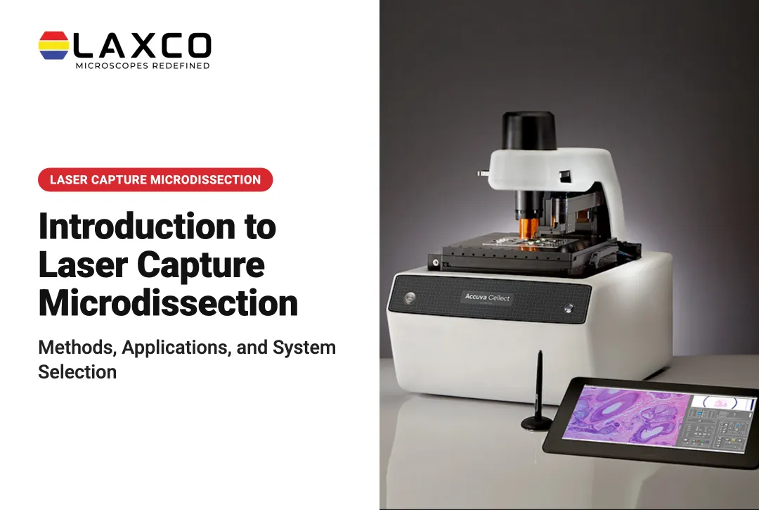
Laxco's Accuva Cellect Laser Capture Microdissection System is designed for this upstream isolation step:
- An infrared (IR) laser designed for film capture of single cells or small numbers of cells.
- An ultraviolet (UV) laser designed for cutting dense structures, such as bone or plant tissue.
- A five-step software workflow with visual quality control and documentation.
- Documented support for FFPE and fresh-frozen samples, several staining approaches, and cell culture.
Laxco presents protein extraction and mass spectrometry among the downstream workflows in its microgenomics workflow. The downstream analysis itself is performed by the researcher's laboratory, not by Laxco.
Common Challenges and Best Practices
Common Sources of Error
- Detergent contamination: trace detergents can suppress ionization.
- Polyethylene glycol (PEG) contamination: a documented source of interfering peaks in LC-MS.
- Incomplete digestion: leaves missed cleavages that complicate peptide quantification.
- Excessive processing: each additional transfer step can increase analyte loss.
Best Practices
- Use MS-grade solvents and reagents.
- Minimize transfer and processing steps.
- Validate with matrix-matched controls, not only neat standards.
- Document each protocol step to support reproducibility across runs and operators.
Frequently Asked Questions
Which preparation method suits plasma samples?
Protein precipitation is commonly used for plasma because it is simple and fast. Where phospholipids cause matrix effects, SPE, SLE, or phospholipid-removal plates may be more suitable. The choice depends on the analyte and required sensitivity.
Can laser capture microdissection samples be used for proteomics?
Yes, LCM-isolated material can be used for protein analysis by mass spectrometry. The small amounts obtained require low-input extraction and sensitive LC-MS/MS methods. Pilot studies help establish how much tissue area is needed for detection.
Which sample types does Accuva Cellect support?
Laxco lists FFPE and fresh-frozen tissue, H&E-stained and LCM-optimized stained sections, immunofluorescence, unstained samples, and cell culture. Confirm compatibility with your extraction and MS protocol during workflow planning.
Can poor sample preparation be corrected after the MS run?
Generally not. Ion suppression, contamination, and analyte loss cannot be fully corrected computationally after acquisition. Internal standards can compensate for some variability, but careful preparation remains the main control.