Key Takeaways
- Organoid analysis combines imaging, molecular profiling, and computational methods.
- Techniques include brightfield and confocal imaging, immunostaining, single-cell sequencing, and AI-assisted segmentation.
- Consistent imaging and validated segmentation reduce variability across large studies.
- Single-organoid measurements can reveal subpopulations that well-level averages hide.
What Is Organoid Analysis?
Organoids are cell-based models that recreate many structural and functional aspects of organs (Zhao et al., Nature Reviews Methods Primers, 2022). Organoid analysis characterizes these models using three broad approaches:
- Morphological: size, count, shape, and structural descriptors.
- Molecular: genomic, transcriptomic, and protein profiling.
- Functional: viability, apoptosis, and drug-response measurements.
Research applications include:
- Drug screening: testing compound effects on viability and growth.
- Disease modeling: studying cancer, developmental disorders, and genetic conditions.
- Developmental biology: studying tissue formation and differentiation.
- Biobanking: cataloging patient-derived organoid lines.
- Toxicology: assessing compound effects at the preclinical stage.
Why Rigorous Analysis Matters
As organoid experiments scale to multiwell screening formats, measurement consistency becomes as important as culture conditions. Researchers must account for differences in organoid number, size, growth rate, plating density, imaging conditions, and well-to-well variability.
For screening assays, the Z′ factor is commonly used to evaluate the separation between positive and negative control responses relative to their variability. Signal-to-background measurements, control-well variation, and reproducibility across independent experiments provide additional information about assay quality.
These metrics must be interpreted in the context of the specific organoid model, endpoint, and experimental design. A satisfactory plate-quality metric does not independently establish biological validity or confirm that an imaging system accurately measures individual organoids.
A review of image-based phenotypic screening with organoids discusses the importance of appropriate controls, replication, and reproducible image-based measurements.
Rigorous analysis:
- Improves the accuracy of size, count, and viability measurements.
- Reduces observer bias and variability between users.
- Increases throughput in screening studies.
- Supports long-term tracking of growth and differentiation.
Single-organoid measurements add further information. Using single-organoid interferometry, Tebon and colleagues found resistant subpopulations ranging from 7.8% to 87.1%, depending on cell line and drug (Nature Communications, 2023). A well-level average would not show this variation.
A Stepwise Organoid Analysis Workflow
Common early errors include inconsistent imaging planes, unvalidated segmentation thresholds, and ignoring the Z-axis in 3D structures.

Step 1: Define the Research Objective
Decide whether the study requires morphological quantification, molecular profiling, or drug-response measurement. This choice determines the imaging modality and analysis software.
Step 2: Capture Images or Samples
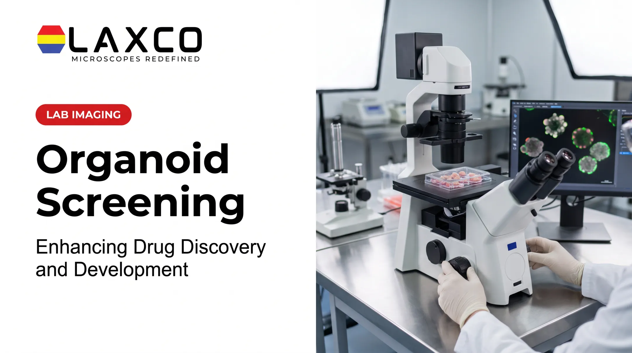
Use brightfield, confocal, or high-content imaging for morphology. For molecular work, organoids may be dissociated into single cells, or fixed and sectioned. Image resolution and sample integrity at this stage directly affect reproducibility.
Step 3: Determine Whether Targeted Tissue Isolation Is Needed (Optional)
For routine organoid imaging, size measurement, counting, or image-based drug-response analysis, tissue isolation is generally unnecessary. Researchers can proceed directly from image acquisition to quantitative image analysis.
When the research question requires molecular profiling of a specific organoid structure or cell population, the organoid may be appropriately fixed or frozen, sectioned, and prepared for laser capture microdissection (LCM).
LCM allows selected regions from compatible organoid sections to be isolated for subsequent DNA, RNA, or protein analysis. The preparation and collection workflow must be validated for the intended molecular assay.
For studies requiring this additional step, Laxco's Accuva Cellect Laser Capture Microdissection System provides:
- An infrared (IR) laser designed for film capture of single cells or small numbers of cells.
- An ultraviolet (UV) laser designed for cutting dense structures.
- A five-step software workflow with visual quality control and documentation.
Step 4: Apply Quantitative or Molecular Analysis
Run automated segmentation and counting, or molecular pipelines such as scRNA-seq or spatial transcriptomics. The chosen methods determine throughput and whether rare populations can be detected.
Step 5: Interpret Results
Relate size, count, and expression data to growth kinetics, dose-response curves, or heterogeneity patterns. Adequate replication and statistical power support confident conclusions.
Step 6: Refine Protocols
Use findings to adjust culture conditions, drug concentrations, or imaging schedules for the next round. Organoid studies are usually iterative.
Example: Testing a Compound's Effect on Growth
- Define the objective: measure growth inhibition across treatment groups.
- Capture baseline images: brightfield imaging across a multi-well plate records starting size and count for each organoid.
- Investigate unusual morphology: identify organoids showing atypical size, shape, or growth patterns after treatment. These findings may justify additional investigation but do not, by themselves, demonstrate drug resistance. Where appropriate, selected organoids can undergo further imaging, molecular profiling, or functional testing.
- Quantify changes: use validated image analysis to compare organoid number, projected size, growth, and other relevant measurements across treatment groups. When organoids overlap or boundaries are unclear, confirm segmentation accuracy and use additional imaging, manual review, or predefined exclusion criteria where necessary.

Common mistakes:
- Using different imaging planes at baseline and endpoint.
- Interpreting unusual morphology or a single imaging endpoint as evidence of drug resistance without suitable follow-up validation.
- Trusting automated counts before validating segmentation thresholds.
The results inform whether to continue with the compound and how to adjust dose range, timing, or imaging frequency.
Laxco Tools for Organoid Analysis
Image analysis. Laxco's LEAP AI pairs Accuva LEAP optics with Media Cybernetics Image-Pro AI:
- Pre-trained deep-learning models for AI-assisted segmentation and measurement.
- An Image → Process → Measure → Report workflow.
- Batch-processable life-science protocols with no-code options.
Live imaging. Laxco's CellLive systems operate inside CO₂ incubators, allowing researchers to monitor cultures over extended experiments without repeatedly removing them from their controlled environment. Images are acquired at scheduled intervals rather than through uninterrupted video recording.
The CellLive CL11 supports brightfield time-lapse monitoring, while the CL22 adds phase contrast, multichannel fluorescence, and Z-stack scanning for more advanced imaging requirements.
Region isolation. Laxco's Accuva Cellect Laser Capture Microdissection System can isolate selected cells or regions from appropriately prepared organoid sections for downstream molecular analysis. Laxco describes the system as having an approximately 32% smaller footprint than comparable microscope-and-laser systems, helping laboratories conserve bench space.
Support. Laxco provides application training and technical support for its microscopy and imaging systems. For Accuva Cellect, Laxco specifically documents training and a 48-hour support response time. Support arrangements for LEAP AI and CellLive should be confirmed for the relevant product and service agreement.
The Cellect Grant Assistance Program provides Grant Language Kits for NIH, NSF, HHMI, and DOD proposals that include LCM.
Protocols that work in a 24-well pilot often need adjustment at 384-well scale. Revalidate imaging and segmentation settings as the study grows.
Frequently Asked Questions
Which sample types does Accuva Cellect support for organoid work?
Laxco lists FFPE and fresh-frozen samples, H&E-stained and LCM-optimized stained sections, immunofluorescence, unstained samples, and cell culture. Organoids are typically fixed or frozen and sectioned before microdissection.
How is organoid size and count measured?
Manual counting under the microscope remains common but is slow and varies between observers. Automated segmentation sizes and counts organoids more consistently across plates, once thresholds are validated against manual annotation.
Can AI segmentation be used for organoid images?
Yes. AI-assisted segmentation is widely used in organoid research. LEAP AI uses pre-trained deep-learning models for measurement, and results should be validated for each organoid type and imaging mode.
Why is single-organoid or single-cell analysis useful?
Well-level measurements average across organoids and cells, which can obscure minority populations. Single-organoid imaging and single-cell sequencing reveal heterogeneity, rare cell types, and differing drug responses.