Key Takeaways
- Fluorescence microscopes combine filtered excitation and emission light with fluorophore labels to image specific targets.
- Widefield, confocal, multiphoton, and TIRF systems differ in depth, resolution, and live-sample suitability.
- System choice depends on sample type, required resolution, number of fluorophores, and budget.
- Image-analysis software influences how quickly and consistently fluorescence data can be quantified.
What Is a Fluorescence Microscope?
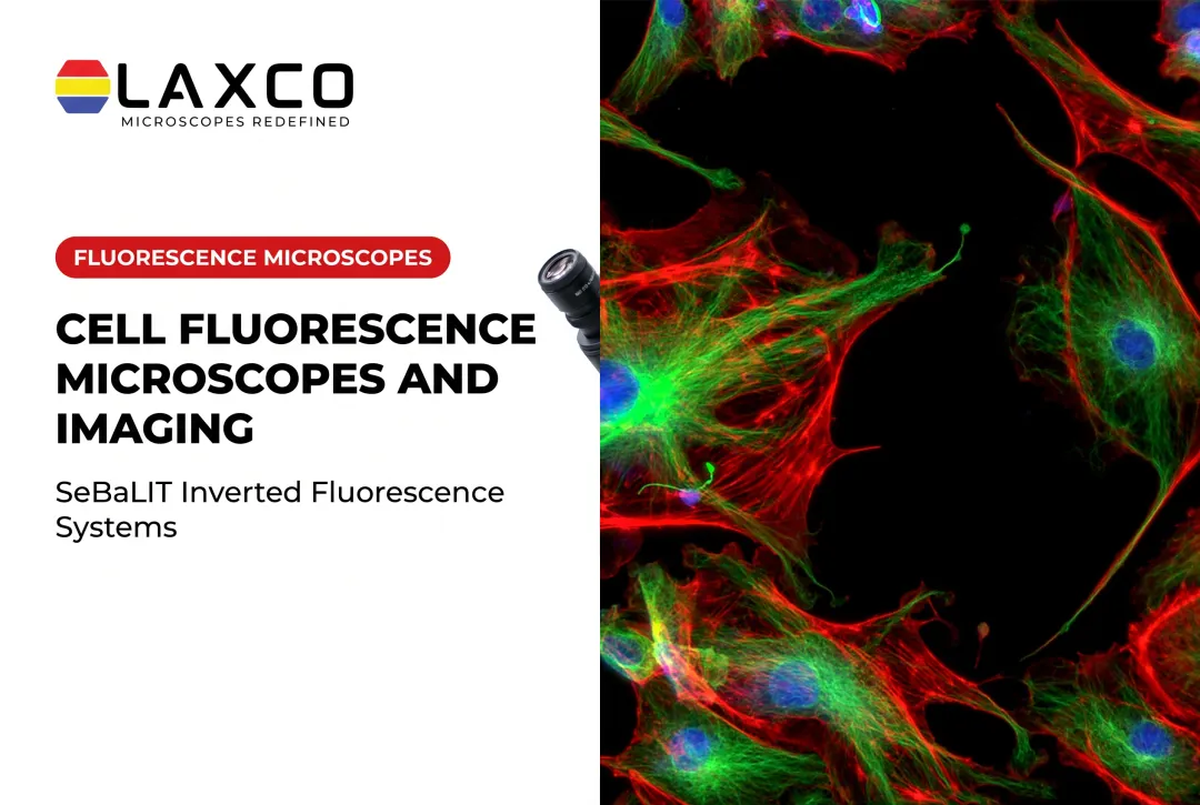
A fluorescence microscope uses fluorophores attached to specific targets to make those targets visible against a dark background. Unlabeled material contributes little signal.
Brightfield microscopy forms images from transmitted light. Fluorescence relies on a wavelength shift between absorbed and emitted light, known as the Stokes shift.
Three optical components separate the signal:
- An excitation filter selects the illumination band.
- A dichroic mirror directs excitation light to the sample and passes emitted light.
- An emission filter passes only the desired signal to the detector.
Common research uses include:
- Protein localization in cell biology.
- Immunology and cancer research.
- Identification of specific cell populations in tissue.
- Target-engagement assays in drug discovery.
Why Fluorescence Microscopy Is Widely Used
Brightfield imaging shows structure. Fluorescence addresses more specific questions, such as whether a protein is expressed in a location or whether a cell is alive. Low-abundance targets that are hard to see in brightfield can be detected when labeled with a bright fluorophore.
Tuberculosis screening provides a documented example of fluorescence microscopy's benefits in a specific diagnostic application. A 2011 World Health Organization policy statement concluded that LED fluorescence microscopy was more sensitive than conventional Ziehl-Neelsen staining for detecting acid-fast bacilli in tuberculosis screening.
In a separate 2019 study at a district hospital in Ghana, researchers evaluated 200 sputum samples from 100 patients with suspected pulmonary tuberculosis. Using Xpert MTB/RIF as the reference test, fluorescence microscopy achieved 84.5% sensitivity compared with 54.8% for Ziehl-Neelsen staining. Average slide-reading time was approximately 1.5 minutes with fluorescence versus 4.6 minutes with Ziehl-Neelsen staining.
These results describe the staining methods, patient samples, and experimental conditions evaluated. They should not be interpreted as universal performance figures for all fluorescence microscopes or as evidence of clinical diagnostic validation for Laxco's SLiPro research instruments.

In research labs, image analysis is often the slowest step. Laxco's LEAP AI pairs Accuva LEAP optics with Media Cybernetics Image-Pro AI for AI-assisted segmentation and measurement. Pre-trained models are intended to reduce variability between users once validated for the sample type.
Types of Fluorescence Microscopes
Widefield (Epifluorescence) Microscopy
Widefield systems illuminate the whole field of view at once, using the same objective for excitation and collection.
- Commonly used for: routine cell biology, immunofluorescence, and teaching.
- Strengths: fast acquisition, lower cost, and a wide field of view.
- Limitation: out-of-focus light reduces contrast in thicker samples.
A review reported widefield lateral resolution of about 200 nm, with good temporal resolution but no optical sectioning (PMC). Widefield imaging is commonly used for thin, fixed samples.
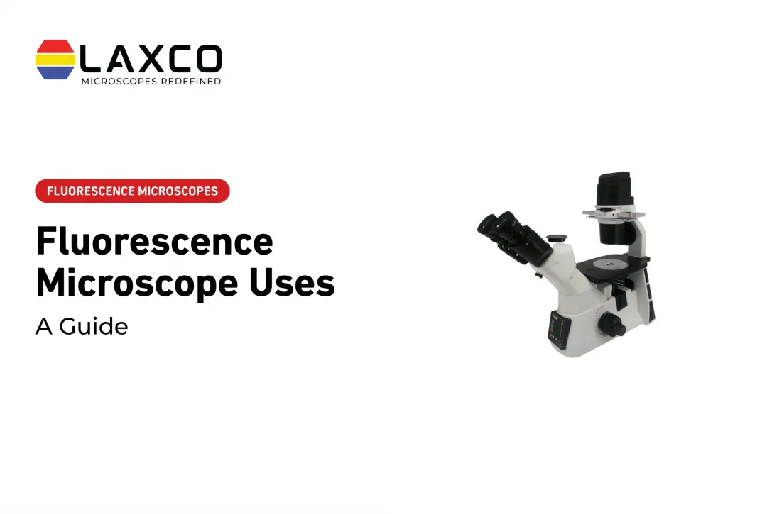
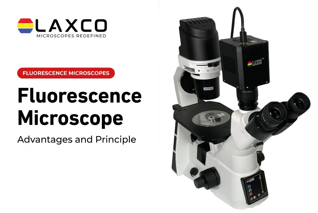
Laxco's inverted widefield fluorescence microscopes include:
- The SLi3Pro, which uses patented SeBaLIT™ illumination and supports DAPI, FITC, and TRITC fluorochromes.
- The SLi6Pro, an inverted widefield fluorescence microscope using SeBaLIT™ illumination, supports three-color multiplex fluorescence imaging across an expanded spectral range of approximately 390-662 nm. It is compatible with more than 110 fluorochromes, including CFP, YFP, and RFP. The 110-plus figure describes the range of supported dyes, not simultaneous imaging of 110 markers.
Confocal Microscopy
Confocal microscopy uses focused illumination and a detection pinhole to reject out-of-focus fluorescence. In conventional laser-scanning confocal systems, a focused beam scans across the sample to build an image. This allows optical sections to be collected at different depths for three-dimensional reconstruction.
Commonly used for: protein localization, detailed cellular structures, thicker specimens, and optical sectioning.
Strengths: rejection of out-of-focus light, improved image contrast in thicker specimens, and depth-resolved imaging. Resolution depends on the optical configuration rather than being automatically superior to widefield microscopy in every dimension.
Limitations: greater instrument complexity and cost. Point-scanning systems may have longer acquisition times and increased photobleaching risk, depending on illumination and imaging settings.
A 2020 review of confocal microscopy describes achievable resolution approaching approximately 0.2 µm laterally and 0.6 µm axially in suitable configurations. These are representative high-performance values, not guaranteed specifications for every confocal microscope. Objective numerical aperture, wavelength, pinhole size, and detection conditions affect actual resolution.
Confocal microscopy is particularly useful when out-of-focus light limits the interpretability of widefield images or when optical sections are required.
Multiphoton Microscopy
Multiphoton systems use pulsed infrared light. Excitation requires near-simultaneous absorption of two or more photons, which occurs mainly at the focal plane.
- Commonly used for: in vivo imaging, live tissue, and deep imaging in neuroscience.
- Strengths: deeper penetration and reduced out-of-focus photodamage.
- Limitation: high cost and slower acquisition than widefield imaging.
A 2017 review of deep-tissue multiphoton microscopy noted that many conventional two-photon studies achieved imaging depths around 500 µm, particularly in living brain tissue. This is a representative depth for the imaging conditions discussed, not a fixed maximum or a universal capability of every multiphoton system.
Achievable depth depends on tissue scattering and absorption, excitation wavelength, fluorophore properties, optical design, and laser power. Specialized systems can image deeper under suitable conditions, although signal loss, photodamage, and tissue heating remain important practical constraints.
Total Internal Reflection Fluorescence (TIRF) Microscopy
TIRF uses an evanescent field to illuminate a thin layer, typically tens to a few hundred nanometers, next to the coverslip.
- Commonly used for: membrane dynamics, single-molecule studies, and receptor trafficking.
- Strengths: very low background and high axial selectivity.
- Limitation: only structures near the coverslip are imaged.

How to Choose a Fluorescence Microscope
Factors to consider:
- Sample type: fixed tissue slides are commonly examined with upright microscopes, while cells growing in culture vessels often benefit from inverted microscope configurations. Widefield, confocal, and multiphoton microscopy describe imaging methods rather than upright or inverted instrument orientation. Select the imaging method according to specimen thickness, required optical sectioning, imaging depth, and live-cell requirements.
- Resolution and sectioning: routine screening rarely needs optical sectioning, whereas 3D structural work does.
- Fluorophore panel: match the number and spectra of labels to the system's illumination and filters.
- Expandability: confirm that filters, cameras, and analysis software can be added later.
- Analysis workload: assess the image-acquisition, segmentation, measurement, batch-processing, and reporting capabilities actually included with the selected microscope and software configuration. Advanced image-analysis platforms may be separate purchases and should not be assumed to be integrated with every fluorescence microscope.
Laxco's LEAP AI pairs Accuva LEAP optics with Media Cybernetics Image-Pro AI for AI-assisted segmentation, measurement, batch processing, and reporting. The workflow follows Image → Process → Measure → Report and supports configurable no-code protocols once models are validated for the sample type. LEAP AI is a separate analysis platform and is not built into the SLi3Pro, SLi6Pro, or CellLive instruments.
For incubator-based live-cell imaging, Laxco's CellLive series is model-specific:
- CL11: primarily brightfield monitoring for confluence, morphology, and time-lapse imaging.
- CL22: adds phase contrast and multichannel fluorescence with Z-stack and quantitative capability. Evaluate CL22 when incubator-based fluorescence imaging is required.
A hands-on evaluation with your own samples remains the most reliable comparison. Assess signal-to-background ratio, exposure requirements, supported fluorescence channels, and the analysis steps each system's software handles. Confirm compatibility across the multi-platform configuration you intend to use.
Frequently Asked Questions
What are the main types of fluorescence microscopy?
The main types are widefield, confocal, multiphoton, and TIRF. Each suits a different combination of resolution, imaging depth, and sample type. Super-resolution methods extend resolution beyond the diffraction limit for specialized applications.
Which Laxco microscope suits three-color fluorescence?
The SLi3Pro is an inverted widefield microscope supporting DAPI, FITC, and TRITC fluorochromes with SeBaLIT™ illumination. It is field-upgradeable, so fluorescence and digital imaging can be added later.
Which Laxco microscope suits larger fluorophore panels?
The SLi6Pro supports three-color multiplex fluorescence imaging across an expanded spectral range of approximately 390-662 nm and is compatible with more than 110 fluorochromes, including CFP, YFP, and RFP, but that compatibility figure does not mean 110 markers can be imaged simultaneously.
Share your fluorophore list with a Laxco specialist so illumination, filters, and detection can be confirmed for your panel.
How can photobleaching be reduced?
Use the lowest illumination intensity and shortest exposure that give adequate signal. Acquire only the channels and time points needed, and use photostable dyes where possible.