Key Takeaways
- Fixing tissue before dissociation can reduce stress-related expression changes caused by enzymatic digestion.
- Alcohol and aldehyde fixatives affect RNA differently, and platform compatibility varies.
- Probe-based hybridization chemistries are designed to work with fragmented RNA from fixed samples.
- Single-nucleus approaches are commonly used for formalin-fixed paraffin-embedded (FFPE) tissue, but they change which cell types are recovered.
- Morphology checks, RNA quality metrics, and small pilot runs reduce the risk of failed libraries.
Why Fix Cells Before scRNA-seq?
Enzymatic dissociation at physiological temperature can induce immediate-early genes, such as FOS, JUN, and EGR1, and heat-shock responses. These changes can be mistaken for biological signals. Dissociation can also reduce recovery of fragile cell types.
Fixing tissue before or during dissociation helps preserve the transcriptional state at the time of collection. Fixation also offers practical benefits:
- Validated fixation and storage workflows can enable samples to be stored or shipped while limiting ongoing transcriptional changes; RNA quality still depends on the fixative, storage conditions, duration, and downstream assay.
- Timepoints or collection sites can be processed together, which reduces batch effects.
- Validated fixation protocols can reduce or eliminate infectivity for some biological agents, but pathogen inactivation depends on the organism, fixative, concentration, exposure time, and biosafety protocol. Fixation should not be assumed to guarantee inactivation unless the procedure has been validated for the specific hazard.
Choosing a Fixation Method
Alcohol and Aldehyde Fixation
For fresh cell suspensions, methanol (an alcohol fixative) and paraformaldehyde (PFA, an aldehyde fixative) are commonly used. Methanol dehydrates and precipitates proteins. PFA forms crosslinks. Each affects RNA recovery and library complexity differently, so the choice should follow the library chemistry being used.
FFPE Tissue
Formalin fixation crosslinks RNA and, over time, fragments it. Conventional poly(dT) capture relies on intact poly(A) tails and can perform poorly on archived FFPE material.
Probe-Based Chemistry
Probe-based methods hybridize short probes to target transcripts rather than capturing poly(A) tails. Because they target short RNA regions, they tolerate fragmentation better. Platform documentation should be checked for supported fixatives and sample types before fixing irreplaceable material.
Fixed-Cell and Single-Nucleus Approaches
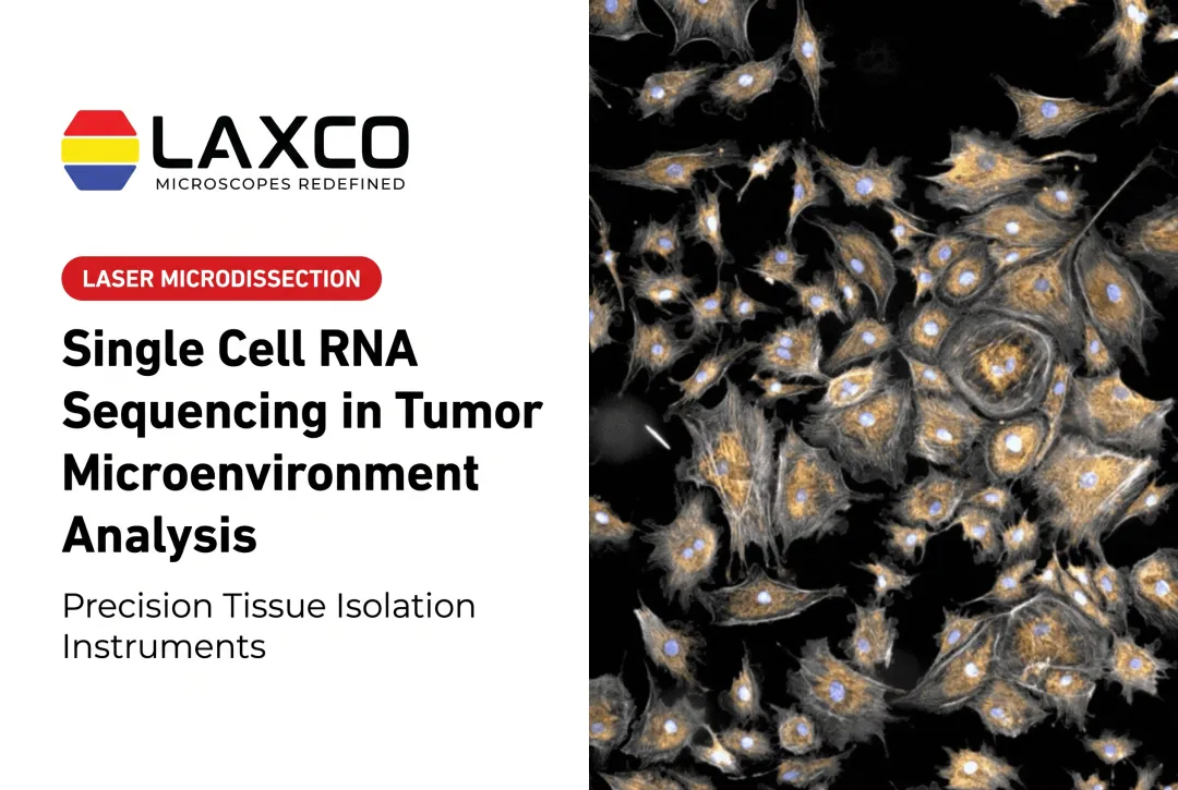
FFPE tissue requires specialized fixed-sample workflows. Depending on the platform and preparation, profiling may use dissociated fixed cells or isolated nuclei. These approaches can recover different RNA compartments and cell populations, so the method should be matched to the tissue, fixation history, and supported library chemistry. Nuclear RNA differs from whole-cell RNA, and some cell types may be under- or overrepresented in nuclear libraries.
General Workflow for Fixed-Cell scRNA-seq
- Fix under controlled conditions. Avoid under-fixation, which can leave morphology or molecular preservation inadequate, and excessive fixation, which can reduce RNA accessibility or recovery. Store fixed material using the temperature, buffer, and duration specified by the validated platform protocol.
- Optimize dissociation or nuclei isolation. Adjust digestion time and mechanical handling for the tissue and assay. Use the temperature and enzyme system validated for the workflow; cold-active dissociation can reduce stress-induced transcriptional artifacts in compatible protocols. Wide-bore tips and gentle handling can help reduce mechanical damage where appropriate.
- Follow validated hybridization and library protocols. Deviations from tested conditions are a common cause of poor libraries.
- Check morphology before sequencing. Inspect cells or nuclei for integrity, debris, and clumping by microscopy, and record images for reference.
- Run a small pilot. Compare conditions on a limited scale before committing a full sample set.
Quality Control Checkpoints
Integrity and counts:
- Record cell viability before fixation when the workflow requires it.
- After fixation, use platform-appropriate fluorescent nucleic-acid stains or microscopy to count cells or nuclei and assess integrity, debris, and clumping; do not interpret post-fixation staining as conventional live/dead viability.
- Use automated or manual counting methods appropriate to the fixed-sample chemistry for consistent loading.
- Use RNA quality or fragment-size metrics appropriate to the sample and assay; DV200 is commonly used for FFPE RNA, but the relevant threshold depends on the validated library workflow.
Debris and multiplets:
- Filtration or sorting to remove fragments and aggregates
- Microscopic review of the suspension
- Computational doublet detection after sequencing
Region-Specific Analysis From Fixed Sections
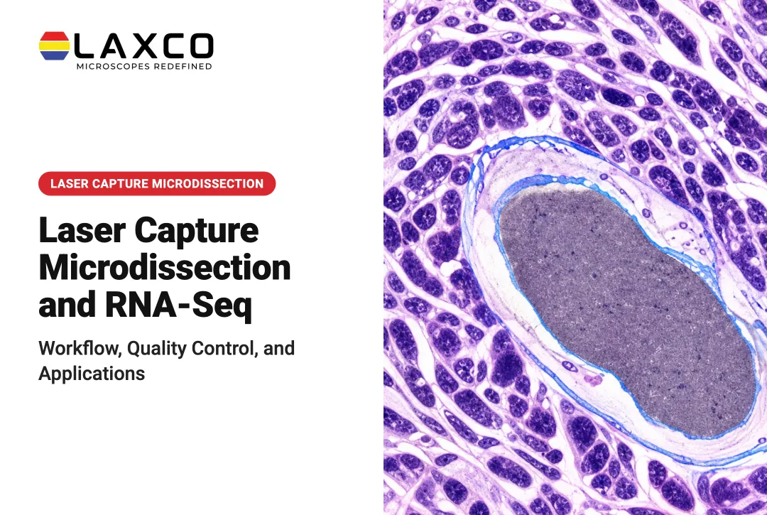
Dissociation-based single-cell workflows lose much of the original spatial context. When the research question concerns a defined region of an FFPE section, such as a tumor area or lesion, laser capture microdissection (LCM) can isolate selected cells or tissue regions while retaining their relationship to morphology.
LCM is complementary to dissociation-based single-cell profiling and should not be assumed to feed directly into a standard scRNA-seq workflow unless the downstream extraction and library method has been specifically validated for the captured material.
A typical LCM workflow loads the slide, defines the target region, confirms the target by visual inspection, and captures the selected cells or region for downstream molecular analysis.

Laxco Systems for Fixed-Sample Workflows
- Accuva Cellect: a laser capture microdissection system with an infrared laser for capture of single cells or small numbers of cells and a UV laser for cutting dense structures. Laxco lists FFPE and fresh-frozen tissue, H&E-stained and LCM-optimized stains, immunofluorescence, unstained samples, and cell culture as supported.
- SeBa digital microscopes: support image capture and documentation, which can be used to record morphology checks of cell or nuclei suspensions.
Laxco supplies instruments with application training and technical support. It does not perform sequencing or library preparation as a laboratory service. Laxco's Cellect Grant Assistance Program provides Grant Language Kits for NIH, NSF, HHMI, and DOD proposals that include Accuva Cellect.
Frequently Asked Questions
Can I use archived FFPE blocks for single-cell studies?
Yes, with adapted methods. Specialized fixed-sample workflows can profile dissociated fixed cells or isolated nuclei from FFPE tissue, often using probe-based chemistries designed to tolerate fragmented RNA. Confirm compatibility with the selected platform, assess sample quality using the metrics recommended for that workflow, and run a small pilot before committing irreplaceable blocks.
When should I use LCM instead of whole-tissue dissociation?
Use LCM when the cells of interest occupy a defined region of a section, or when morphology determines which cells to analyze. Dissociation-based methods suit profiling all cell types in a tissue without location.
How do I check nuclei or cell quality before sequencing?
Inspect suspensions by microscopy for intact nuclei, debris, and clumps, and record images for each sample. Laxco's SeBa digital microscopes integrate camera and display, which simplifies capturing and archiving these morphology checks.
Which sample types does Accuva Cellect support?
Laxco lists FFPE and fresh-frozen tissue, H&E-stained and LCM-optimized stained sections, immunofluorescence, unstained samples, and cell culture. For fixed-tissue RNA work, confirm compatibility with your extraction and library protocols.
What support does Laxco provide for new LCM users?
Laxco lists training and 48-hour response times as part of its Accuva Cellect support offering. Its Cellect Grant Assistance Program provides grant-support resources for qualifying instrumentation proposals.