Key Takeaways
- LCM isolates defined cells or regions under microscopic guidance before liquid chromatography-tandem mass spectrometry (LC-MS/MS).
- Compared with MALDI imaging, LCM-MS gives up a continuous spatial map in exchange for deeper proteome coverage of selected regions.
- Fixation and staining can affect protein and peptide recovery, but the effect depends on the preparation and extraction protocol. Low-input LCM samples require appropriately optimized proteomics workflows.
- At low input, sample handling and surface losses are major sources of variability.
- Formalin-fixed paraffin-embedded (FFPE) archives can be analyzed with adapted LCM-MS workflows.
How LCM and Mass Spectrometry Work Together
LCM uses infrared (IR) or ultraviolet (UV) lasers under direct visualization to collect selected cells or regions from tissue sections. Mass spectrometry, usually LC-MS/MS, then identifies and quantifies peptides from the collected material.
- LCM defines what is analyzed, based on the region the researcher selects.
- Mass spectrometry identifies or provides evidence for proteins through their measured peptides and can estimate relative protein abundance. Absolute quantification requires an appropriately calibrated analytical method.
In this workflow, spatial resolution is set by the microdissection boundary rather than by the mass spectrometer.
LCM-MS Compared With MALDI Imaging
MALDI imaging mass spectrometry measures molecules directly across a tissue section and preserves a continuous spatial map. In the 2013 Maier et al. study, matrix extraction plus complementary LC-MS/MS identified about 1,400 proteins across ten human tissues; that figure is not direct MALDI image identification of 1,400 spatially localized proteins (Maier et al., 2013).
LCM with LC-MS/MS is region-based, not a continuous pixel map. It analyzes a defined microdissected region by solution-based LC-MS/MS, which can provide greater proteome depth for that region.

The LCM-MS Workflow
1. Sectioning and Staining
Tissue is cut into thin sections and mounted on slides compatible with the microdissection system. H&E, immunohistochemistry, or other suitable staining methods may help identify target regions. Effects on protein and peptide recovery depend on the stain, fixation, tissue, and extraction procedure. Choose a staining protocol that provides reliable target identification and is validated for the intended downstream proteomics workflow.
2. Microdissection and Collection
The researcher defines the target region, and the laser captures or cuts it. The collected material is transferred to a cap or tube for extraction. Each transfer at low input can cause losses, so handling steps should be kept to a minimum.
3. Lysis, Retrieval, and Digestion
This stage typically includes:
- Protein solubilization from the captured material.
- FFPE protein recovery: heat-assisted treatment and suitable extraction chemistry can improve protein solubilization and partially reverse formalin-induced crosslinks, although fixation-related modifications may remain.
- Enzymatic digestion of proteins into peptides.
Because input amounts are small, minor inconsistencies at this stage can affect results.
4. Chromatography and Mass Spectrometry
Peptides are separated by low-flow liquid chromatography and analyzed by tandem mass spectrometry. Methods designed for bulk tissue may need adjustment for the small loads produced by microdissection.
A Published Example
A 2020 study described an LCM-MS method for H&E-stained FFPE human lung tissue (Herrera et al., 2020). The authors noted that fixation and H&E staining lower protein yield with standard preparation methods. They used heat extraction, physical disruption, and in-column digestion to analyze microdissected alveolar regions.
Applications
Cancer Research
LCM-MS can separate malignant cells from stroma or immune cells and compare their proteomes. This supports studies of tumor heterogeneity, signaling pathways, and candidate therapeutic targets.
Developmental Biology
Developing tissues contain distinct compartments that change over time. Microdissection allows each compartment to be profiled separately across developmental stages.
Drug Discovery
Cell-type-specific proteome data can show whether a target is expressed in the intended compartment and how a compound affects specific regions of a tissue.
Archival FFPE Tissue
FFPE tissue makes up most clinical archives. Adapted LCM-MS workflows allow retrospective studies that link proteomic findings to existing clinical records.
Challenges and Limitations
- Fixation and staining can affect protein recovery. Some preparation methods reduce extraction efficiency or peptide identification, while optimized workflows can mitigate these effects. Validate extraction and digestion for the specific tissue, fixation, and staining protocol.
- Small samples magnify losses. Contamination and adsorption to tube surfaces matter more at low input. Low-binding consumables and fewer transfers help.
- Throughput depends on target size and workflow. Manual region identification and microdissection can be time-consuming, particularly when substantial tissue input is required. Processing time varies by sample complexity, required capture area, operator experience, and instrument capabilities.
- Region selection shapes the result. Selection criteria should be documented as part of the method.
Choosing an LCM System for Proteomics
When comparing LCM systems for mass spectrometry workflows, consider:
- Laser type and collection mechanism: both IR film-based capture and UV cutting can isolate small targets, depending on the platform. Compare target size, tissue compatibility, capture efficiency, collection method, and downstream requirements. Confirm support for dense structures where relevant.
- Sample compatibility: FFPE, fresh-frozen, stained, immunofluorescence, and unstained sections.
- Collection mechanism: how captured material is transferred with minimal loss.
- Software and documentation: how target selection and quality control are recorded.
- Training and support: availability of application training and response times.
Laxco Accuva Cellect for LCM-MS
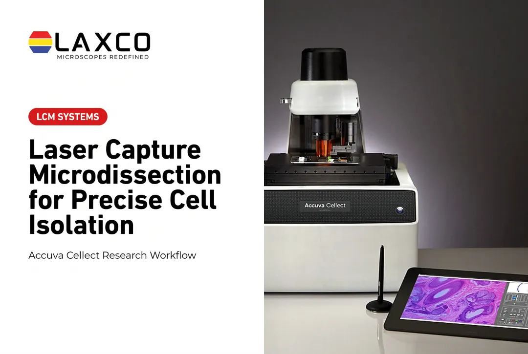
Laxco's Accuva Cellect Laser Capture Microdissection System enables researchers to isolate selected cells or tissue regions before downstream protein extraction and mass-spectrometry analysis. The instrument handles tissue selection and collection, while protein extraction, digestion, and mass spectrometry are separate analytical steps. Its documented features include:
- Infrared (IR) laser: designed for capture of single cells or small numbers of cells.
- UV laser: designed for cutting dense structures, such as bone or plant tissue.
- Sample preparation: Laxco lists FFPE and fresh-frozen tissue, H&E-stained and LCM-optimized stains, immunofluorescence, unstained samples, and cell culture.
- Collection: compatible with CapSure LCM caps.
- Software workflow: a five-step workflow with visual quality control.
Laxco lists training and 48-hour response times for Accuva Cellect. Accuva Cellect supports the tissue-isolation stage of research workflows; mass-spectrometry analysis is performed separately by the research laboratory or core facility. Laxco's Cellect Grant Assistance Program provides Grant Language Kits for NIH, NSF, HHMI, and DOD proposals.
Frequently Asked Questions
How much tissue do I need for LCM-MS proteomics?
Requirements depend on tissue type, fixation, staining, and the sensitivity of your mass spectrometer. Run a pilot with several capture areas to find the smallest input that gives adequate protein identifications before collecting study samples.
Does H&E staining reduce protein recovery?
Effects are protocol-specific. A 2020 study of H&E-stained FFPE human lung tissue reported that fixation and H&E staining can lower protein yield with standard preparation methods, and used adapted protocols (heat extraction, physical disruption, and in-column digestion) to analyze microdissected alveolar regions (Herrera et al., 2020). Those findings describe that workflow and should not be generalized to all H&E or FFPE preparations.
Can Accuva Cellect collect material for mass spectrometry?
Yes. Accuva Cellect isolates selected cells or tissue regions; it does not perform mass spectrometry. Protein extraction, digestion, and MS analysis are separate steps carried out by the research laboratory or core facility. Confirm that your lysis buffer and digestion protocol are compatible with the collection cap before starting a study.
Which tissue preparations can I microdissect for proteomics?
Laxco lists FFPE and fresh-frozen tissue, H&E-stained and LCM-optimized stained sections, immunofluorescence, unstained samples, and cell culture for Accuva Cellect. Staining does not produce a uniform reduction in protein recovery; effects depend on the stain, fixation, tissue, and extraction protocol. Validate the preparation with your intended proteomics workflow.
How does Accuva Cellect handle dense tissue for proteomics?
Accuva Cellect includes a UV laser designed for cutting dense structures, such as bone or plant tissue, alongside an infrared laser for single cells or small groups. This allows one system to sample both cell populations and larger regions.