Key Takeaways
- scRNA-seq has refined the classical hematopoietic hierarchy by revealing continuous transcriptional transitions, lineage priming, and heterogeneity within conventionally defined progenitor populations.
- Transcriptional differences among HSCs can reflect cell-cycle state as well as differences in fate.
- Transcriptomic clusters suggest candidate cell states that can be tested and validated with lineage tracing, transplantation, or other functional assays.
- In acute myeloid leukemia (AML), scRNA-seq has been used to resolve malignant cell states that vary between patients.
- Cell viability, sorting conditions, and batch design strongly affect single-cell data quality.
From a Lineage Tree to a Continuum
The classical model of hematopoiesis proceeds from HSCs to multipotent progenitors (MPPs), lineage-committed progenitors, and mature cells. Population-level assays measure the average of each sorted compartment, which can hide variation within it.
Surface Markers and Transcriptional States
Researchers commonly enrich stem and progenitor compartments using surface-marker panels before sequencing. In mouse studies, these populations are often defined within a lineage-negative, Sca-1-positive, c-Kit-positive (LSK) compartment. Simplified examples include:
| Compartment | Example mouse phenotype |
|---|---|
| Long-term HSC (LT-HSC) | Lin− Sca-1+ c-Kit+ CD34− CD135/Flt3− |
| Short-term HSC (ST-HSC) | Lin− Sca-1+ c-Kit+ CD34+ CD135/Flt3− |
| Flt3+ MPP subset | Lin− Sca-1+ c-Kit+ CD34+ CD135/Flt3+ |
These are simplified example phenotypes rather than universal definitions. HSC and MPP gating varies across studies, mouse strains, and marker panels, and SLAM-family markers such as CD150 and CD48 are also commonly used. Single-cell data further show that cells within one immunophenotypic gate can occupy different transcriptional states.
A Continuous Differentiation Program
A 2016 study applied scRNA-seq to zebrafish hematopoietic cells undergoing thrombocyte commitment (Macaulay et al., 2016). By ordering cells computationally, the authors placed each cell along a continuum from stem cell to mature cell. Progression along this continuum involved coordinated suppression of proliferation and ribosomal biogenesis genes as lineage-specific genes increased.
Interpreting HSC Heterogeneity
LT-HSC populations show transcriptional heterogeneity. Some of this variation reflects cell-cycle state rather than differences in self-renewal or lineage fate.
- Clonal barcoding methods link individual HSC clones to their later progeny, providing functional context for transcriptional states.
- Without lineage tracing or transplantation data, a transcriptional cluster is a hypothesis about fate.
- Combining transcriptomic and functional data gives a more reliable assignment of stem cell states.
Transcript Isoforms
Short-read sequencing is well suited to counting transcripts but provides limited information about full-length isoforms. Long-read sequencing can resolve splice variants. Studies combining both approaches in mouse HSCs and progenitors have described isoform diversity that gene-level counts do not capture.
Applications in Blood Cancer Research
Malignant Cell States in AML
A 2019 study in Cell profiled 38,410 cells from bone marrow aspirates of patients with AML and healthy donors (van Galen et al., 2019). The authors classified malignant cells along a hierarchy related to normal hematopoiesis. Primitive AML cells often co-expressed stemness and myeloid-priming programs, a pattern the authors did not observe in normal cells.
Drug Response by Cell Population
Single-cell approaches allow drug responses to be measured in defined subpopulations rather than as an average across a sample. This can show whether a compound affects malignant cells, normal progenitors, or both.
Biomarker Research
Single-cell studies in AML have reported cell-state features associated with relapse or remission. These associations are exploratory biomarker findings and require validation in independent cohorts before clinical use.
Technical Considerations
Common challenges include:
- Low cell numbers: rare compartments such as LT-HSCs yield limited starting material.
- Batch effects: samples processed in different runs can differ for technical reasons.
- Handling stress: sorting, dissociation, and temperature changes can alter expression before sequencing.
- Doublets and ambient RNA: both can create artificial intermediate states.

Preserving Marrow Architecture
Most hematopoiesis scRNA-seq studies use dissociated marrow or blood. Questions about the bone marrow niche require spatial information. Laser capture microdissection (LCM) can isolate selected cells or regions from marrow sections, so molecular data can be related to location, such as proximity to vessels or the endosteum.
Laxco Tools for Hematopoiesis Research
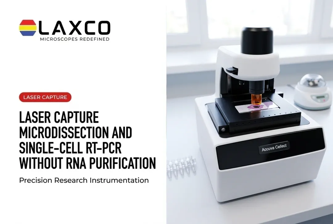
- Accuva Cellect: a laser capture microdissection system. Its infrared laser captures single cells or small numbers of cells, and its UV laser cuts dense structures, including bone. Laxco lists FFPE and fresh-frozen tissue, several staining types, immunofluorescence, unstained samples, and cell culture as supported.
- LEAP AI: combines Accuva LEAP optics with Media Cybernetics Image-Pro AI for image processing, AI-assisted segmentation, measurement, and reporting through an Image → Process → Measure → Report workflow. LEAP AI is a separate microscopy and image-analysis platform from Accuva Cellect.
For Accuva Cellect, Laxco lists training and 48-hour response times. Laxco provides instruments and related support; it does not perform sequencing as a laboratory service.
Frequently Asked Questions
Can Accuva Cellect work with bone marrow sections?
Accuva Cellect includes a UV laser designed for cutting dense structures, including bone. Decalcification and fixation affect RNA quality, so confirm that your section preparation is compatible with downstream sequencing before collecting study samples.
Should I use FACS or LCM for hematopoietic cells?
FACS suits isolating cells from marrow aspirates or blood by surface markers. LCM suits isolating cells or regions from marrow sections when location in the niche, such as proximity to vessels or bone, matters.
How can I study the bone marrow niche spatially?
Combine imaging of marrow sections with region-specific isolation. LCM can collect cells near defined structures for sequencing, while multiplexed imaging maps protein markers. Both approaches retain information that dissociation removes.
Can image analysis help characterize marrow sections?
LEAP AI combines Accuva LEAP optics with Media Cybernetics Image-Pro AI for AI-assisted segmentation, measurement, image processing, and reporting. It can support image-based characterization of marrow sections, but Laxco does not document LEAP AI as an automated target-selection integration for Accuva Cellect.
What training is available for new Accuva Cellect users?
Laxco lists training and 48-hour response times as part of its Accuva Cellect support offering. Confirm the available training format and application-specific support directly with Laxco when planning the system for marrow or bone-section workflows.